 Indietro
Chapter 16- Lecture 4
Guida di studio - Note intelligenti
 Appunti personalizzati basati sui tuoi materiali, ampliati con definizioni chiave, esempi e contesto.
The Autonomic Nervous System (ANS)
Overview of the ANS
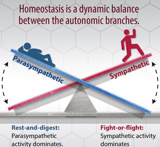
The autonomic nervous system is a division of the peripheral nervous system responsible for regulating involuntary physiological functions, including heart rate, blood vessel diameter, digestion, and respiratory rate. It consists of two main branches: the sympathetic and parasympathetic divisions, which often have opposing effects on target organs.
Sympathetic Division: Prepares the body for 'fight or flight' responses.
Parasympathetic Division: Promotes 'rest and digest' activities.
Dual Innervation: Most vital organs receive input from both divisions, allowing fine-tuned regulation.
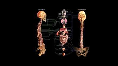
Dual Innervation
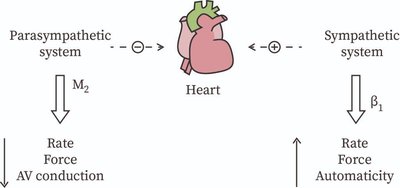
Dual innervation refers to the phenomenon where most organs are innervated by both sympathetic and parasympathetic fibers, which typically exert opposing effects. This arrangement allows for precise control of organ function and homeostasis.
Autonomic Plexuses: Networks of nerves formed by mingled sympathetic postganglionic and parasympathetic preganglionic fibers, especially in the thoracic and abdominopelvic cavities.
Autonomic Tone: The background level of activity in autonomic motor neurons, which can be increased or decreased for greater control.
Example: The heart receives dual innervation; parasympathetic activity dominates at rest, while sympathetic activity increases during crisis.
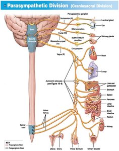
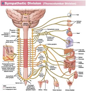
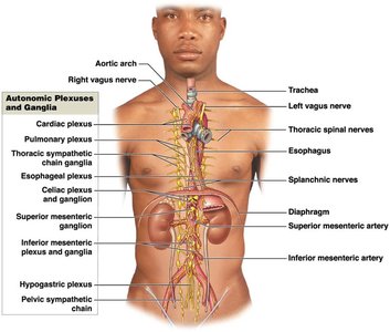
Single Innervation and Autonomic Tone
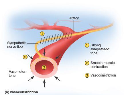
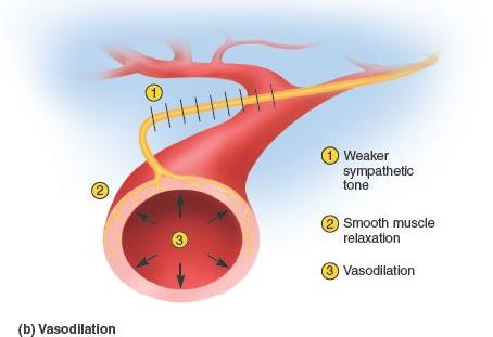
Some organs are innervated by only one division of the ANS. For example, blood vessel diameter is primarily controlled by the sympathetic division.
Sympathetic Tone: Maintains partial contraction of smooth muscle in blood vessel walls, allowing for rapid adjustments in vessel diameter.
Vasoconstriction: Increased sympathetic activity causes smooth muscle contraction and vessel narrowing.
Vasodilation: Decreased sympathetic activity causes smooth muscle relaxation and vessel widening.
Hierarchy of ANS Control
Levels of Control in the ANS
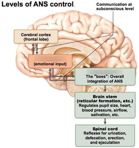
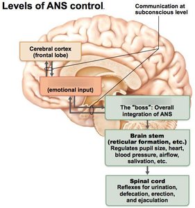
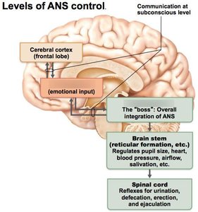
The autonomic nervous system is regulated by a hierarchy of control centers within the central nervous system, from the cerebral cortex to the spinal cord.
Cerebral Cortex: Modifies ANS activity at a subconscious level, often through the limbic system.
Hypothalamus: Integrates visceral, emotional, and behavioral responses; acts as the main control center for homeostasis.
Brainstem (Medulla Oblongata): Controls moment-to-moment involuntary processes such as heart rate and respiration.
Spinal Cord: Mediates reflexes for urination, defecation, erection, and ejaculation.
Voluntary Control of Visceral Functions
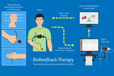
While most autonomic functions are involuntary, some can be influenced consciously through techniques such as biofeedback therapy, which trains individuals to control certain physiological processes.
Biofeedback Therapy: Uses real-time feedback to help patients learn to control heart rate, blood pressure, and other functions.
Visceral Reflexes
Structure and Function of Visceral Reflexes
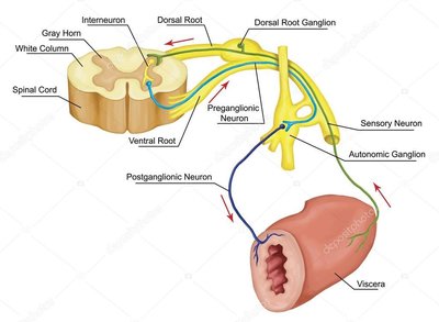
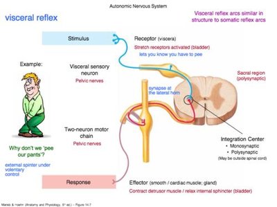
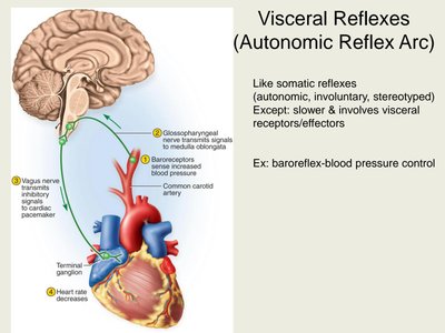
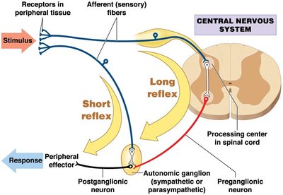
Visceral reflexes are autonomic, polysynaptic reflexes initiated in the internal organs (viscera) to maintain homeostasis. They provide automatic motor responses to internal stimuli.
Components of a Visceral Reflex Arc:
Receptor
Sensory neuron
Processing center (interneurons)
Two sequential visceral motor neurons
Effector (smooth muscle, cardiac muscle, or gland)
Example: Bladder stretch receptors initiate a reflex arc to control urination.
Long vs. Short Visceral Reflexes
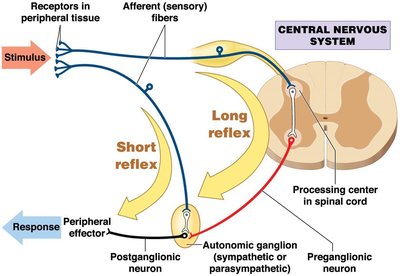
Visceral reflexes are classified as long or short based on their pathways and complexity.
Long Reflexes:
Involve the central nervous system (CNS).
Slower and more complex, often integrating emotional or external stimuli.
Example: Emotional stress affecting heart rate.
Short Reflexes:
Bypass the CNS, with sensory neurons directly stimulating postganglionic neurons in local nerve plexuses.
Faster and more immediate responses.
Example: Stomach contraction in response to food stretching the wall.
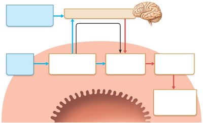
Neural Reflex Pathways in the Gastrointestinal Tract
The gastrointestinal tract is regulated by both long and short reflexes, allowing for independent control of digestive functions.
External Stimuli: Sight, smell, taste, or thought of food can initiate long reflexes via the CNS.
Internal Stimuli: Chemoreceptors, osmoreceptors, and mechanoreceptors in the GI tract initiate short reflexes within local nerve plexuses.
Effectors: Smooth muscle or glands respond by changing contractile or secretory activity.
Summary Table: Comparison of Long and Short Visceral Reflexes
Feature | Long Reflex | Short Reflex |
|---|---|---|
Pathway | Involves CNS | Bypasses CNS |
Speed | Slower | Faster |
Complexity | Complex, integrates emotions/external stimuli | Simple, local response |
Example | Emotional stress affecting heart rate | Stomach contraction to digest food |
Key Terms and Definitions
Dual Innervation: The supply of an organ by both sympathetic and parasympathetic fibers.
Autonomic Tone: The baseline level of activity in autonomic motor neurons.
Visceral Reflex: An automatic response to internal stimuli, mediated by the ANS.
Autonomic Plexus: A network of autonomic nerves serving a region of the body.
Homeostasis: The maintenance of a stable internal environment.
Relevant Equations
Blood Pressure Regulation (Baroreflex): Where is the change in pressure, is blood flow, and is resistance.
Conclusion
The autonomic nervous system is essential for maintaining internal homeostasis through dual innervation, autonomic tone, and visceral reflexes. Understanding the hierarchy of control and the distinction between long and short reflexes is crucial for comprehending how the body responds to internal and external stimuli. Additional info: Academic context was added to clarify the structure and function of visceral reflexes, the hierarchy of ANS control, and the physiological significance of dual innervation and tone.
