 Indietro
The Autonomic Nervous System: Structure, Function, and Clinical Relevance
Guida di studio - Note intelligenti
 Appunti personalizzati basati sui tuoi materiali, ampliati con definizioni chiave, esempi e contesto.
Autonomic Nervous System (ANS) Overview
Introduction to the ANS
The autonomic nervous system (ANS) is a division of the peripheral nervous system responsible for regulating involuntary physiological processes, including heart rate, blood pressure, respiration, digestion, and glandular activity. It operates largely below the level of consciousness and is essential for maintaining homeostasis.
Effectors: Smooth muscle, cardiac muscle, and glands
Functions: Adjusts blood flow, heart rate, blood pressure, and digestive processes
Control: Subconscious (involuntary)
Alternate names: Involuntary nervous system, visceral motor system

Organization of the Nervous System
Central and Peripheral Divisions
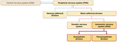
The nervous system is divided into the central nervous system (CNS) and the peripheral nervous system (PNS). The PNS is further divided into sensory (afferent) and motor (efferent) divisions. The motor division includes the somatic nervous system (voluntary control of skeletal muscles) and the autonomic nervous system (involuntary control of visceral effectors).

ANS vs. Somatic Nervous System
Key Differences
Although both systems have motor fibers, they differ in their effectors, neurotransmitters, and neural pathways.
Effectors: Somatic system targets skeletal muscle; ANS targets cardiac muscle, smooth muscle, and glands.
Neurotransmitters: Somatic neurons release acetylcholine (ACh), always excitatory. ANS preganglionic fibers release ACh; postganglionic fibers release ACh or norepinephrine (NE), which can be excitatory or inhibitory.
Neural Pathways: Somatic system uses a single neuron from CNS to effector; ANS uses a two-neuron chain (preganglionic and postganglionic neurons).

Divisions of the Autonomic Nervous System
Parasympathetic and Sympathetic Divisions
The ANS is divided into two main branches with generally opposing effects:
Parasympathetic Division: Promotes maintenance functions and conserves energy ("rest and digest").
Sympathetic Division: Mobilizes the body during activity ("fight or flight").

Parasympathetic Division
The parasympathetic division maintains body functions during restful situations.
Blood pressure, heart rate, and respiratory rate are low
Gastrointestinal tract activity is high
Pupils constricted, lenses accommodated for close vision
Example: Relaxing and reading after a meal

Sympathetic Division
The sympathetic division prepares the body for stressful or energetic activity.
Increases heart rate, dilates pupils, causes cold, sweaty skin
Shunts blood to skeletal muscles and heart
Dilates bronchioles, causes liver to release glucose
Activated by exercise, excitement, emergency, embarrassment

Dual Innervation and Homeostasis
Dynamic Antagonism
Most visceral organs receive input from both sympathetic and parasympathetic divisions, which usually have opposite effects. This dynamic antagonism maintains homeostasis.
Example: Sympathetic division increases heart rate; parasympathetic division decreases it.
Iris of the eye: Sympathetic dilates pupil; parasympathetic constricts pupil (antagonistic effect).
Salivary glands: Sympathetic stimulates mucus secretion; parasympathetic stimulates enzyme secretion (cooperative effect).
Neurotransmitters and Receptors of the ANS
Key Neurotransmitters
The effects of the ANS are determined by the neurotransmitters released and the receptors present on target organs.
Acetylcholine (ACh): Released by all preganglionic neurons and parasympathetic postganglionic neurons (cholinergic).
Norepinephrine (NE): Released by most sympathetic postganglionic neurons (adrenergic).
Anatomical Differences Between Sympathetic and Parasympathetic Divisions
Key Anatomical Features
Sites of Origin: Parasympathetic fibers are craniosacral (brain and sacral spinal cord); sympathetic fibers are thoracolumbar (thoracic and lumbar spinal cord).
Relative Lengths of Fibers: Parasympathetic has long preganglionic and short postganglionic fibers; sympathetic has short preganglionic and long postganglionic fibers.
Location of Ganglia: Parasympathetic ganglia are in or near visceral effectors; sympathetic ganglia are close to the spinal cord.
Visceral Reflexes
Structure and Function
Visceral reflexes control involuntary functions such as digestion and blood pressure. They share basic components with somatic reflex arcs but have key differences.
All reflex arcs begin with sensory input
Components: receptor, sensory neuron, integration center, motor neuron, effector
Differences from somatic reflexes:
Two consecutive neurons in the motor pathway
Afferent fibers are visceral sensory neurons
Effectors are smooth muscle, cardiac muscle, and glands
Examples: Reflexes that empty the rectum and bladder; baroreflex for blood pressure regulation
Neural Pathways: Somatic vs. Autonomic
Pathway Structure
Somatic Pathway: Single motor neuron from CNS to skeletal muscle
Autonomic Pathway: Two-neuron chain (preganglionic and postganglionic) from CNS to effector
Clinical Considerations and Disorders of the ANS
Common Disorders
Autonomic neuropathy: Damage to autonomic nerves, often due to diabetes mellitus. Symptoms include sexual dysfunction, dizziness, urinary incontinence, sluggish pupil reactions, and impaired sweating. Prevention focuses on good blood glucose control.
Hypertension: Overactive sympathetic vasoconstrictor response to stress, leading to high blood pressure. Treated with adrenergic receptor-blocking drugs (beta blockers).
Raynaud’s disease: Exaggerated vasoconstriction in fingers and toes, causing them to turn pale or cyanotic. Treated with vasodilators.
Autonomic dysreflexia: Uncontrolled activation of autonomic neurons in individuals with spinal cord injuries, causing dangerous increases in blood pressure and risk of stroke.
Control of ANS Activity
ANS is regulated by CNS centers: brainstem, spinal cord, hypothalamus, and cerebral cortex
Hypothalamus is the main integrative center
Cerebral input can modify ANS activity subconsciously via the limbic system
Summary Table: Comparison of Somatic and Autonomic Motor Neurons
Feature | Somatic Nervous System | Autonomic Nervous System |
|---|---|---|
Effectors | Skeletal muscle | Smooth muscle, cardiac muscle, glands |
Neural Pathway | Single neuron from CNS to effector | Two-neuron chain (preganglionic and postganglionic) |
Neurotransmitter at Effector | Acetylcholine (ACh) | ACh or Norepinephrine (NE) |
Effect | Always excitatory | Excitatory or inhibitory |
Additional info: The enteric nervous system, a subdivision of the ANS, is involved in gastrointestinal reflexes and is composed of sensory neurons, interneurons, and motor neurons located in the walls of the GI tract.
