 Indietro
The Axial and Appendicular Skeleton: Structure, Function, and Clinical Relevance
Guida di studio - Note intelligenti
 Appunti personalizzati basati sui tuoi materiali, ampliati con definizioni chiave, esempi e contesto.
The Human Skeleton: Overview
Divisions and Functions
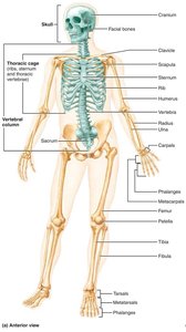
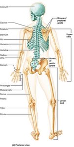
The human skeleton is a structural framework composed of bones, cartilages, joints, and ligaments. It is divided into two major regions: the axial skeleton and the appendicular skeleton. The skeleton provides support, protection, and facilitates movement.
Axial skeleton: Includes the skull, vertebral column, and thoracic cage. Forms the longitudinal axis of the body and protects vital organs.
Appendicular skeleton: Includes the limbs and girdles, enabling movement and manipulation of the environment.
Functions: Support, protection, movement, mineral storage, and blood cell formation.
The Axial Skeleton
Skull: Structure and Function
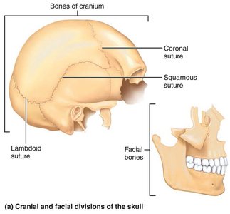
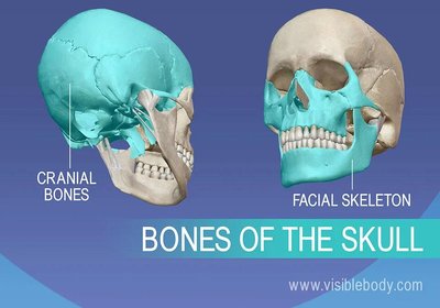
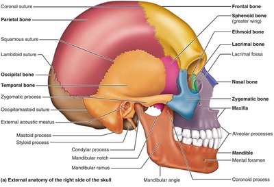
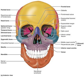
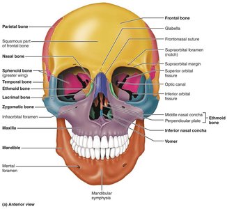
The skull is composed of cranial and facial bones, which protect the brain and form the framework of the face.
Cranial bones: Enclose the brain and provide attachment sites for head and neck muscles.
Facial bones: Form the face, house sensory organs, provide openings for air and food, secure teeth, and anchor facial muscles.
Sutures: Immovable joints connecting skull bones, with a serrated appearance.
Skull Geography and Cavities
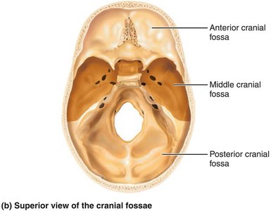
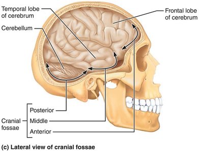
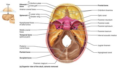
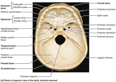
The cranium is divided into a vault and a base, with internal fossae supporting the brain. The skull contains multiple cavities and openings for nerves and blood vessels.
Cranial vault (calvaria): Forms the superior, lateral, and posterior portions of the skull.
Cranial base: Forms the inferior aspect, divided into anterior, middle, and posterior cranial fossae.
Cavities: Middle/internal ear, nasal cavity, orbits, and sinuses.
Openings: Foramina, canals, and fissures for passage of nerves and vessels.
Bony Boundaries of Orbits and Nasal Cavity
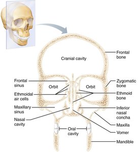
The orbits and nasal cavity are formed by multiple bones, providing protection and passageways for sensory organs.
Orbits: Roof (frontal, sphenoid), lateral wall (zygomatic, sphenoid), floor (maxilla, zygomatic, palatine), medial wall (ethmoid, lacrimal, maxilla, sphenoid).
Nasal cavity: Roof (nasal, frontal, ethmoid, sphenoid), floor (maxilla, palatine), lateral walls (maxilla, ethmoid, inferior nasal conchae, palatine), medial wall (ethmoid, vomer).
Paranasal sinuses: Frontal, ethmoidal, sphenoidal, and maxillary sinuses lighten the skull and amplify voice.
Skull Bones and Markings
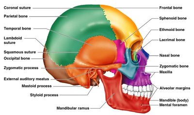
The skull consists of 22 bones, with distinct markings for muscle attachment and passage of nerves and vessels.
Cranial bones: Frontal, parietal, occipital, temporal, sphenoid, ethmoid.
Facial bones: Mandible, maxillae, zygomatic, nasal, lacrimal, palatine, vomer, inferior nasal conchae.
Markings: Foramina (openings), processes (projections), sutures (joints).
The Vertebral Column
Structure and Regions
The vertebral column extends from the skull to the pelvis, transmitting weight and protecting the spinal cord. It consists of 33 vertebrae in five regions.
Cervical: 7 vertebrae (neck)
Thoracic: 12 vertebrae (upper back)
Lumbar: 5 vertebrae (lower back)
Sacrum: 5 fused vertebrae (pelvis)
Coccyx: 4 fused vertebrae (tailbone)
Curvatures and Functions
The vertebral column exhibits four natural curvatures, contributing to shock absorption and flexibility.
Cervical and lumbar: Convex anteriorly
Thoracic and sacral: Concave anteriorly
Functions: Shock absorption, balance, flexibility
Structure of a Typical Vertebra
Body: Main weight-bearing structure
Vertebral arch: Formed by pedicles and laminae
Processes: Spinous, transverse, superior/inferior articular
Regional Features
Cervical: Smallest, transverse foramina, bifid spinous processes
Thoracic: Articular facets for ribs, heart-shaped body, long spinous processes
Lumbar: Largest, kidney-shaped body, short blunt spinous processes
The Thoracic Cage
Structure and Function
The thoracic cage protects vital organs and supports the shoulder girdle. It consists of the sternum, ribs, and thoracic vertebrae.
Sternum: Manubrium, body, xiphoid process
Ribs: True ribs (1-7), false ribs (8-12), floating ribs (11-12)
Functions: Protection, support, respiration
True vs. False Ribs
Type | Attachment | Connection |
|---|---|---|
True Ribs (1-7) | Direct | Own costal cartilage |
False Ribs (8-12) | Indirect | Shared or no connection |
Floating Ribs (11-12) | None | Do not attach to sternum |
The Appendicular Skeleton
Pectoral Girdle and Upper Limb
The pectoral girdle consists of the clavicle and scapula, allowing free movement of the upper limb.
Clavicle: Collarbone, fractures anteriorly to protect arteries
Scapula: Shoulder blade, triangular with three borders
Upper limb bones: Humerus (arm), radius and ulna (forearm), carpals (wrist), metacarpals (palm), phalanges (fingers)
Pelvic Girdle and Lower Limb
The pelvic girdle is composed of fused bones (ilium, ischium, pubis) and supports the weight of the upper body.
Hip bones: Ilium, ischium, pubis
Femur: Thigh bone, largest and strongest
Patella: Knee cap, protects and improves leverage
Tibia and fibula: Leg bones, tibia bears weight, fibula does not
Foot: Tarsals (ankle), metatarsals (sole), phalanges (toes), arches support weight
Joints: Classification and Structure
Functional and Structural Classification
Joints are classified by their function and structure, determining their mobility.
Functional: Synarthroses (immovable), amphiarthroses (slightly movable), diarthroses (freely movable)
Structural: Fibrous, cartilaginous, synovial
Fibrous Joints
Suture: Only in skull, immovable
Syndesmosis: Bones connected by ligaments, variable movement
Gomphosis: Peg-in-socket, e.g., teeth
Cartilaginous Joints
Synchondrosis: Hyaline cartilage, immovable
Symphysis: Fibrocartilage, slightly movable (e.g., pubic symphysis, intervertebral joints)
Synovial Joints
Features: Articular cartilage, joint cavity, synovial fluid, fibrous capsule, synovial membrane, ligaments
Bursae: Sacs filled with synovial fluid, reduce friction
Tendon sheath: Elongated bursa around tendons
Factors Affecting Stability
Shape of articular surfaces
Number and positioning of ligaments
Muscle tone
Types of Synovial Joints
Classified by shape: plane, hinge, pivot, condyloid, saddle, ball-and-socket
Clinical Relevance: Bone Health and Disorders
Osteoporosis and Bone Density
Risk factors: Age, genetics, lifestyle, decreased estrogen in postmenopausal women
Prevention: Calcium, vitamin D, weight-bearing exercise, avoiding smoking/alcohol
Screening: DEXA scan for bone density
Treatments: Medications (bisphosphonates, SERMs, hormone therapy)
Rehabilitation and Therapy
Physical and occupational therapy restore function after fractures
Exercise guidelines: Weight-bearing, resistance, flexibility
Joint Disorders
Bursitis: Inflammation of bursae
Tendonitis: Inflammation of tendon sheaths
Arthritis: Over 100 types, including osteoarthritis (aging), rheumatoid arthritis (autoimmune)
Summary Table: Axial vs. Appendicular Skeleton
Region | Main Components | Functions |
|---|---|---|
Axial Skeleton | Skull, vertebral column, thoracic cage | Support, protection, attachment |
Appendicular Skeleton | Limbs, pectoral and pelvic girdles | Movement, manipulation |
