 Indietro
The Axial and Appendicular Skeleton: Structure and Function
Guida di studio - Note intelligenti
 Appunti personalizzati basati sui tuoi materiali, ampliati con definizioni chiave, esempi e contesto.
The Human Skeleton: Overview
Divisions and Functions
The human skeleton is a structural framework composed of bones, cartilages, joints, and ligaments. It is divided into two major regions: the axial skeleton and the appendicular skeleton. The skeleton provides support, protection, and facilitates movement.
Axial Skeleton: Consists of the skull, vertebral column, and thoracic cage. Forms the longitudinal axis of the body and protects vital organs.
Appendicular Skeleton: Includes the limbs and girdles, enabling movement and interaction with the environment.
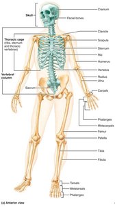
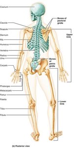
The Axial Skeleton
Skull: Structure and Function
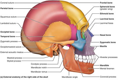
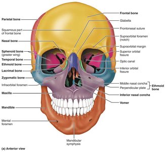
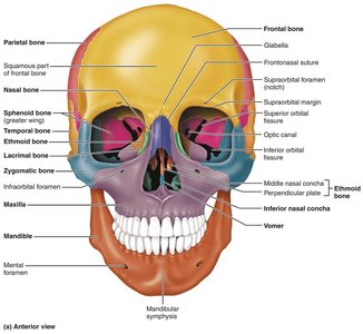
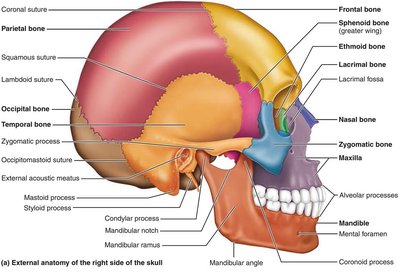
The skull is composed of cranial and facial bones, which protect the brain and form the structure of the face.
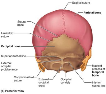
Cranial Bones: Enclose the brain and provide attachment sites for head and neck muscles.
Facial Bones: Form the framework of the face, house sensory organ cavities, and anchor muscles for facial expression.
Sutures: Immovable joints connecting skull bones, with a serrated appearance.
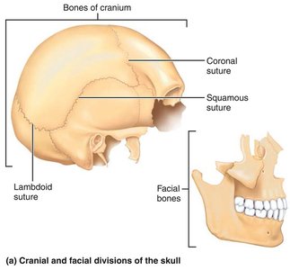
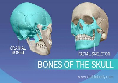
Skull Geography and Cavities
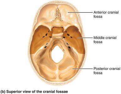
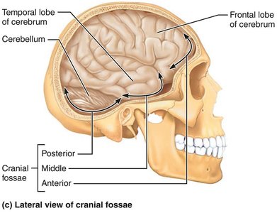
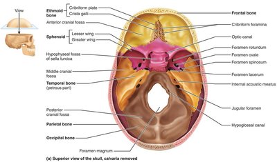
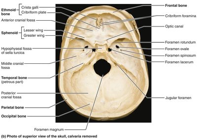
The cranium is divided into a vault and a base, with internal fossae supporting the brain. The skull contains multiple cavities for sensory organs and passageways for nerves and blood vessels.
Cranial Vault (Calvaria): Forms the superior, lateral, and posterior portions of the skull.
Cranial Base: Forms the inferior aspect, divided into anterior, middle, and posterior cranial fossae.
Openings: Foramina, canals, and fissures allow passage of nerves and vessels.
Bony Boundaries of Orbits and Nasal Cavity
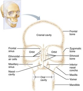
The orbits and nasal cavity are formed by multiple bones, providing protection and support for sensory organs.
Orbits: Roof (frontal, sphenoid), lateral wall (zygomatic, sphenoid), floor (maxilla, zygomatic, palatine), medial wall (ethmoid, lacrimal, maxilla, sphenoid).
Nasal Cavity: Roof (nasal, frontal, ethmoid, sphenoid), floor (maxilla, palatine), lateral walls (maxilla, ethmoid, inferior nasal conchae, palatine), medial wall (ethmoid, vomer).
Paranasal Sinuses: Frontal, ethmoidal, sphenoidal, and maxillary sinuses lighten the skull and amplify voice.
Skull Bones: Markings and Identification
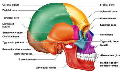
Skull bones have distinct markings for muscle attachment, passage of nerves and vessels, and articulation.
Foramina: Openings for blood vessels and nerves.
Processes: Projections for muscle attachment.
Sutures: Immovable joints connecting skull bones.
The Vertebral Column
Structure and Regions
The vertebral column extends from the skull to the pelvis, transmitting weight and protecting the spinal cord. It consists of 26 irregular bones in five major regions.
Cervical: 7 vertebrae (neck)
Thoracic: 12 vertebrae (upper back)
Lumbar: 5 vertebrae (lower back)
Sacrum: 5 fused vertebrae (pelvis)
Coccyx: 4 fused vertebrae (tailbone)
Curvatures and Functions
The vertebral column exhibits natural curvatures that enhance shock absorption and flexibility.
Cervical and Lumbar: Convex anteriorly
Thoracic and Sacral: Concave anteriorly
Intervertebral Discs: Cushion vertebrae and allow movement
Abnormal Spinal Curvatures
Scoliosis: Abnormal lateral rotation, may cause breathing difficulties
Kyphosis: Excessive thoracic curvature (hunchback)
Lordosis: Excessive lumbar curvature (swayback)
Structure of a Typical Vertebra
Body: Main weight-bearing structure
Vertebral Arch: Formed by pedicles and laminae
Processes: Spinous, transverse, superior and inferior articular processes
Regional Features
Cervical: Smallest, transverse foramina, bifid spinous processes
Thoracic: Articular facets for ribs, heart-shaped body, long spinous processes
Lumbar: Largest, kidney-shaped body, short spinous processes
The Thoracic Cage
Bones and Functions
The thoracic cage protects vital organs and supports the shoulder girdle.
Sternum: Manubrium, body, xiphoid process
Ribs: True ribs (1-7), false ribs (8-12), floating ribs (11-12)
Thoracic Vertebrae: Articulate with ribs
True vs. False Ribs
True Ribs: Attach directly to sternum via costal cartilage
False Ribs: Attach indirectly or not at all
Floating Ribs: No attachment to sternum
The Appendicular Skeleton
Pectoral Girdle and Upper Limb
The pectoral girdle consists of the clavicle and scapula, allowing free movement of the upper limb.
Clavicle: Collarbone, fractures anteriorly to protect arteries
Scapula: Shoulder blade, triangular with three borders
Humerus: Arm bone, articulates with scapula, radius, and ulna
Ulna: Forms elbow joint, olecranon and coronoid processes
Radius: Major contribution to wrist joint
Hand: Carpals (wrist), metacarpals (palm), phalanges (fingers)
Pelvic Girdle and Lower Limb
The pelvic girdle is composed of fused bones (ilium, ischium, pubis) and supports the weight of the upper body.
Femur: Thigh bone, largest and strongest
Patella: Knee cap, protects knee joint
Tibia: Medial, weight-bearing
Fibula: Lateral, non-weight-bearing
Foot: Tarsals (ankle), metatarsals (sole), phalanges (toes)
Arches of the Foot
Medial and Lateral Longitudinal Arches: Support weight, maintained by bone shape, ligaments, and tendons
Transverse Arch: Distributes weight
Joints: Classification and Structure
Functional and Structural Classification
Joints are classified by their movement and structure.
Functional: Synarthroses (immovable), amphiarthroses (slightly movable), diarthroses (freely movable)
Structural: Fibrous, cartilaginous, synovial
Fibrous Joints
Suture: Only in skull, immovable
Syndesmosis: Bones connected by ligaments
Gomphosis: Peg-in-socket, e.g., teeth
Cartilaginous Joints
Synchondrosis: Hyaline cartilage, immovable
Symphysis: Fibrocartilage, slightly movable (e.g., pubic symphysis, intervertebral joints)
Synovial Joints
Articular Cartilage: Covers bone ends
Joint Capsule: Encloses joint cavity
Synovial Fluid: Lubricates joint
Ligaments: Reinforce joint
Bursae and Tendon Sheaths: Reduce friction
Factors Affecting Synovial Joint Stability
Articular Surface Shape
Ligament Number and Position
Muscle Tone
Inflammatory Conditions
Bursitis: Inflammation of bursae
Tendonitis: Inflammation of tendon sheaths
Arthritis: Over 100 types, including osteoarthritis and rheumatoid arthritis
Bone Health and Clinical Considerations
Importance of Load-Bearing Activities
Mechanical Stress: Stimulates bone deposition and increases density
Physical Activity: Reduces risk of osteoporosis and fractures
Bone Imaging Techniques
X-rays: Detailed images of bone and soft tissues
MRI: Cross-sectional images for complex fractures
CT Scans: Detailed bone structure
Hormones and Bone Health
Estrogen: Promotes bone deposition, inhibits resorption
Testosterone: Increases bone density
Parathyroid Hormone (PTH): Regulates calcium, stimulates bone resorption
Exercise Guidelines and Screening
Weight-Bearing Exercises: Walking, running, weightlifting
Bone Density Screening: DEXA scan for osteoporosis risk
Rehabilitation and Therapy
Occupational Therapy: Regains independence
Physical Therapy: Improves strength and balance
Post-Fracture Rehabilitation: Restores function and mobility
Summary Table: Axial vs. Appendicular Skeleton
Region | Main Components | Primary Functions |
|---|---|---|
Axial Skeleton | Skull, vertebral column, thoracic cage | Support, protection, axis formation |
Appendicular Skeleton | Limbs, pectoral and pelvic girdles | Movement, manipulation, interaction |
Summary Table: Types of Joints
Type | Structure | Movement | Examples |
|---|---|---|---|
Fibrous | Connected by fibrous tissue | Immovable or slightly movable | Sutures, syndesmoses, gomphoses |
Cartilaginous | Connected by cartilage | Immovable or slightly movable | Synchondroses, symphyses |
Synovial | Joint cavity with synovial fluid | Freely movable | Knee, shoulder, hip |
Summary Table: Major Bones of the Skull
Bone | Location | Function |
|---|---|---|
Frontal | Forehead, superior orbit | Protection, muscle attachment |
Parietal | Superior/lateral skull | Protection |
Occipital | Posterior skull | Protection, articulation with vertebrae |
Temporal | Inferolateral skull | Protection, muscle attachment |
Sphenoid | Base of skull | Keystone, articulation |
Ethmoid | Deep skull, nasal cavity | Support, olfactory function |
Mandible | Lower jaw | Chewing, muscle attachment |
Maxilla | Upper jaw | Chewing, support |
