 Indietro
The Axial Skeleton: Structure, Function, and Key Anatomical Features
Guida di studio - Note intelligenti
 Appunti personalizzati basati sui tuoi materiali, ampliati con definizioni chiave, esempi e contesto.
The Skeletal System: Overview
Divisions of the Skeleton
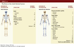
The human skeleton is divided into two main regions: the axial skeleton and the appendicular skeleton. The axial skeleton forms the central supporting axis of the body, while the appendicular skeleton includes the limbs and girdles.
Axial Skeleton: Skull, vertebral column, thoracic cage (ribs and sternum)
Appendicular Skeleton: Upper and lower limbs, pectoral and pelvic girdles
Division of Skeleton | Structure | Number of Bones |
|---|---|---|
Axial Skeleton | Skull (Cranium, Face), Hyoid, Auditory ossicles, Vertebral column, Thoracic cage | 80 |
Appendicular Skeleton | Pectoral girdle, Upper limbs, Pelvic girdle, Lower limbs | 126 |
Anatomical Features of Bones
Bone Surface Markings
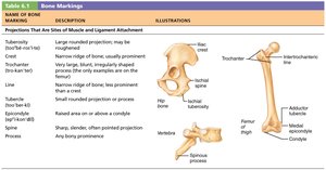
Bones exhibit a variety of markings that serve as sites for muscle attachment, passageways for nerves and blood vessels, and articulation points for joints. Understanding these features is essential for identifying bone functions and relationships.
Projection: Outward bulge of bone, often due to muscle pull or joint modification
Depression: Bowl- or groove-like cut-out, serving as passageways or joint surfaces
Opening: Hole or canal for passage of blood vessels and nerves
Name of Bone Marking | Description | Illustration |
|---|---|---|
Tuberosity | Large rounded projection | Femur, humerus |
Crest | Narrow ridge of bone | Pelvis |
Trochanter | Large, blunt, irregularly shaped process | Femur |
Line | Narrow ridge | Femur |
Tubercle | Small rounded projection | Humerus |
Epicondyle | Raised area above a condyle | Femur |
Spine | Sharp, slender projection | Vertebra |
Process | Any bony prominence | Vertebra |
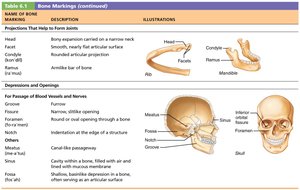
Name of Bone Marking | Description | Illustration |
|---|---|---|
Head | Bony expansion carried on a narrow neck | Femur, humerus |
Facet | Smooth, nearly flat articular surface | Vertebra |
Condyle | Rounded articular projection | Mandible |
Ramus | Arm-like bar of bone | Mandible |
Foramen | Round or oval opening through a bone | Skull |
Meatus | Canal-like passageway | Temporal bone |
Sinus | Cavity within a bone | Skull |
Fossa | Shallow, basin-like depression | Skull |
Groove | Furrow | Skull |
Notch | Indentation at the edge of a structure | Skull |
Axial Skeleton: Structure and Function
Components and Functions
The axial skeleton consists of 80 bones divided into three major regions: the skull, vertebral column, and thoracic cage. It forms the longitudinal axis of the body, supports the head, neck, and trunk, and protects vital organs such as the brain, spinal cord, and thoracic organs.
Skull: Protects the brain and forms the structure of the face
Vertebral Column: Supports the body and protects the spinal cord
Thoracic Cage: Protects the heart and lungs
The Skull
Structure and Sutures
The skull is the most complex part of the skeleton, composed of 22 bones. Most are joined by immovable sutures, which are fibrous joints that appear as seams on the cranial surface. Sutures are synarthroses, allowing minimal movement for compliance and elasticity.
Cranial Bones: Form the cranium, protecting the brain
Facial Bones: Form the structure of the face
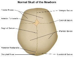
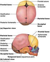
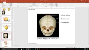
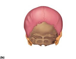
Sutures: Coronal, sagittal, lambdoid, and squamous
Fontanelles in Newborns
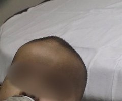
Fontanelles are "soft spots" on the skull of newborns where bones have not yet fused. Premature fusion is known as craniosynostosis. These areas allow for growth and flexibility during birth and early development.
Major Sutures of the Skull
Sagittal Suture: Between parietal bones
Coronal Suture: Separates frontal from parietal bones
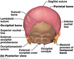
Lambdoid Suture: Joins occipital with parietal bones
Squamous Suture: Joins temporal and parietal bones
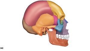
Cranial Bones
Frontal Bone
The frontal bone forms the anterior portion of the cranium, including the forehead. The supraorbital margin is pierced by the supraorbital foramen, allowing passage of the supraorbital artery and nerve. The glabella is the smooth area between the orbits.
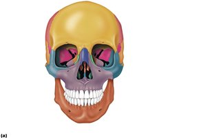
Parietal Bones
The right and left parietal bones form most of the cranial roof and part of its walls. Each is bordered by four sutures that join it to neighboring bones.
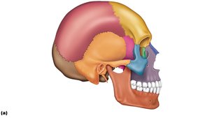
Temporal Bones
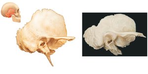
The temporal bones form the lower wall and part of the floor of the cranial cavity. They include the squamous, tympanic, mastoid, and petrous parts, each with distinct features and functions.
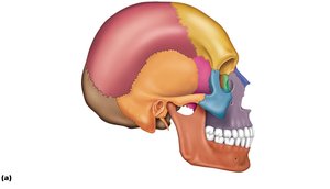
Occipital Bone
The occipital bone forms the rear of the skull and much of its base. Its most prominent feature is the foramen magnum, through which the spinal cord passes. Nuchal lines are sites of muscle attachment.
Sphenoid Bone
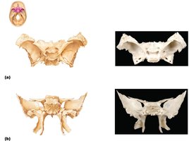
The sphenoid bone is a complex, bat-shaped bone that articulates with all other cranial bones. It contains sphenoidal sinuses and the sella turcica, which houses the pituitary gland. The bone has greater and lesser wings, pterygoid processes, and several foramina for nerve and vessel passage.
Ethmoid Bone
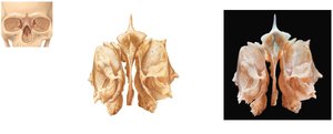
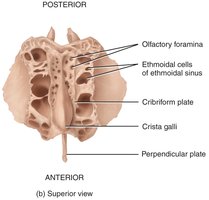
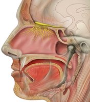
The ethmoid bone is located between the orbital cavities and forms the roof of the nasal cavity. Its perpendicular plate forms the superior part of the nasal septum, and the cribriform plate allows passage of olfactory nerves.
Facial Bones
Overview and Functions
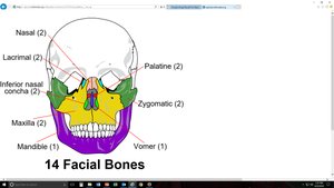
Facial bones do not directly contact the brain. They support the teeth, give shape to the face, form part of the orbital and nasal cavities, and provide attachment for muscles of facial expression and mastication.
Maxillae
The maxillae form the upper jaw and the hard palate, which is the roof of the mouth and the floor of the nasal cavity. Each tooth root is inserted into a deep socket called an alveolus.
Palatine Bones
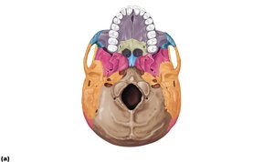
The palatine bones are L-shaped, forming the posterior one-third of the hard palate and part of the nasal cavity and orbits.
Zygomatic Bones
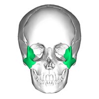
The zygomatic bones form the cheekbones and the inferolateral margins of the orbits. They articulate with the zygomatic processes of the temporal, frontal, and maxillary bones.
Lacrimal Bones
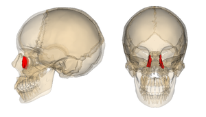
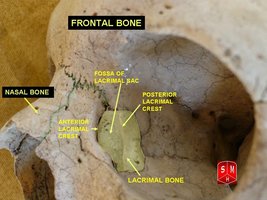
The lacrimal bones are the smallest bones of the skull, forming part of the medial wall of each orbit. The lacrimal fossa houses the lacrimal sac, which drains tears into the nasal cavity.
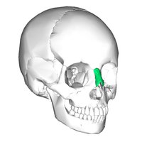
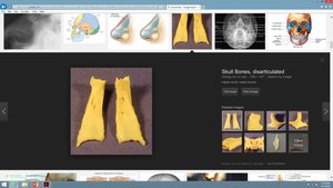
Nasal Bones
The nasal bones form the bridge of the nose and support cartilages that shape the lower portion of the nose.
Nasal Conchae
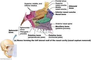
There are three conchae in the nasal cavity: superior and middle (parts of the ethmoid bone), and inferior (a separate facial bone). The conchae increase turbulence of airflow in the nasal cavity.
Vomer
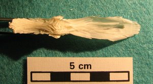
The vomer forms the inferior portion of the nasal septum, separating the left and right nasal cavities. Its name refers to its resemblance to a plow blade.
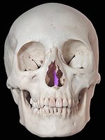
Mandible
The mandible is the largest and strongest bone of the face, forming the lower jaw. It consists of a body (chin) and two upright rami. Key features include the mandibular angle, coronoid process, condylar process, mandibular notch, alveolar process, and foramina for nerves and blood vessels.
Bones Associated with the Skull
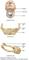
Hyoid Bone
The hyoid bone does not articulate with any other bone. It supports the tongue and provides attachment sites for muscles of the neck and pharynx, helping to keep the larynx open.
Auditory Ossicles
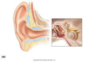
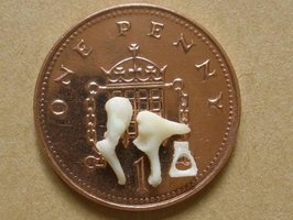
The auditory ossicles (malleus, incus, stapes) are tiny bones in the middle ear associated with hearing.
Orbits and Nasal Cavity
Orbits
The orbits are cavities that encase the eyes and lacrimal glands, serving as sites of attachment for eye muscles. They are formed by parts of seven bones: frontal, sphenoid, zygomatic, maxilla, palatine, lacrimal, and ethmoid.
Nasal Cavity
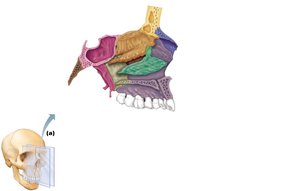
The nasal cavity is formed by several bones. The roof is made by the cribriform plates of the ethmoid, lateral walls by the conchae and perpendicular plates, and the floor by the palatine and maxillary bones. The nasal septum is formed by the vomer and perpendicular plate of the ethmoid, with the anterior portion formed by septal cartilage.
Paranasal Sinuses
Structure and Function
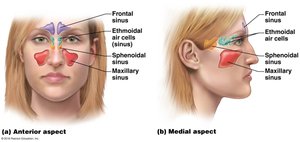
The paranasal sinuses are air cavities situated around the nasal cavity. They help circulate air in the respiratory system and are paired and bilateral. The sinuses include the frontal, ethmoidal, sphenoidal, and maxillary sinuses.
Summary Table: Cranial and Facial Bones
Bone | Number | Function |
|---|---|---|
Frontal | 1 | Forehead, roof of orbits |
Parietal | 2 | Roof and sides of cranium |
Temporal | 2 | Lower sides of cranium, floor |
Occipital | 1 | Back and base of skull |
Sphenoid | 1 | Keystone, articulates with all cranial bones |
Ethmoid | 1 | Roof of nasal cavity, medial wall of orbit |
Maxillae | 2 | Upper jaw, hard palate |
Palatine | 2 | Posterior hard palate |
Zygomatic | 2 | Cheekbones |
Lacrimal | 2 | Medial orbit wall |
Nasal | 2 | Bridge of nose |
Inferior nasal concha | 2 | Largest nasal concha |
Vomer | 1 | Inferior nasal septum |
Mandible | 1 | Lower jaw |
Additional info: Academic context was added to clarify bone functions, anatomical features, and relationships. Tables were reconstructed and expanded for completeness. Images were included only when directly relevant to the explanation of the paragraph.
