 Indietro
The Axial Skeleton: Vertebral Column and Thoracic Cage
Guida di studio - Note intelligenti
 Appunti personalizzati basati sui tuoi materiali, ampliati con definizioni chiave, esempi e contesto.
The Vertebral Column
General Features and Functions
The vertebral column, commonly referred to as the spine, is a flexible, curved structure composed of 26 irregular bones called vertebrae. It physically supports the skull and trunk, allows for their movement, transmits the weight of the trunk to the lower limbs, surrounds and protects the spinal cord, and provides attachment points for ribs and muscles.
Support: Maintains upright posture and supports the head and trunk.
Protection: Encloses and protects the spinal cord.
Movement: Provides flexibility and movement through its articulations.
Attachment: Serves as a site for muscle and rib attachment.
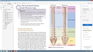
Regions of the Vertebral Column
The vertebral column is divided into five major regions, each with distinct characteristics:
Cervical (C1–C7): 7 vertebrae in the neck region.
Thoracic (T1–T12): 12 vertebrae in the upper back, each articulating with ribs.
Lumbar (L1–L5): 5 vertebrae in the lower back, supporting the most weight.
Sacrum: 1 bone formed from the fusion of 5 vertebrae; articulates with the hip bones.
Coccyx: 1 bone formed from the fusion of 3–5 vertebrae; forms the terminus of the column.
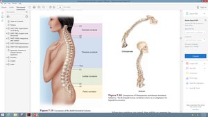
Curvatures of the Vertebral Column
The vertebral column is curved to varying degrees in different locations. These curves increase the strength of the column, help maintain balance, absorb shocks during walking, and protect the vertebrae from fracture.
Cervical and lumbar curves: Concave posteriorly.
Thoracic and sacral curves: Convex posteriorly.
Structure of a Typical Vertebra
All vertebrae share a common structural pattern:
Body (centrum): Anterior, weight-bearing region.
Vertebral arch: Composed of two pedicles and two laminae, forming the posterior part of the vertebra.
Vertebral foramen: Opening formed by the body and arch, through which the spinal cord passes.
Processes: Spinous, transverse, superior and inferior articular processes for muscle and ligament attachment.
Intervertebral foramina: Lateral openings for passage of spinal nerves.
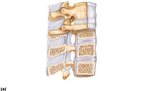
Intervertebral Discs
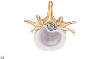
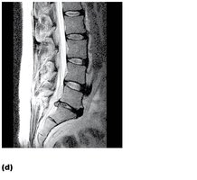
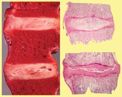
Intervertebral discs are cushion-like pads between vertebrae that act as shock absorbers. Each disc consists of:
Nucleus pulposus: Inner gelatinous core providing elasticity and compressibility.
Anulus fibrosus: Outer collar of collagen and fibrocartilage, limiting expansion of the nucleus pulposus.
Ligaments of the Vertebral Column
Ligaments, along with trunk muscles, help support the vertebral column:
Anterior and posterior longitudinal ligaments: Run along the front and back of the spine, preventing hyperextension and hyperflexion.
Ligamentum flavum: Connects adjacent vertebrae, providing elasticity.
Short ligaments: Connect each vertebra to those above and below.
Abnormal Spinal Curvatures
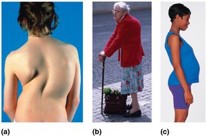
Abnormal curvatures can be congenital or result from disease, poor posture, or muscle imbalance:
Scoliosis: Abnormal lateral rotation, often in the thoracic region; may cause breathing difficulties.
Kyphosis: Excessive dorsal thoracic curvature (hunchback); common in osteoporosis.
Lordosis: Accentuated lumbar curvature (swayback); seen in pregnancy or obesity.
Regional Characteristics of Vertebrae
Cervical Vertebrae (C1–C7)
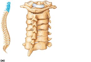
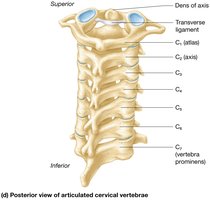
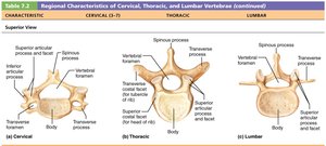
Cervical vertebrae are the smallest and lightest. C3–C7 share these features:
Oval-shaped body
Bifid spinous processes (except C7)
Large, triangular vertebral foramen
Transverse foramen in each transverse process for artery passage
C7 is vertebra prominens, palpable through the skin
Atlas (C1)
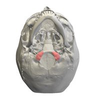
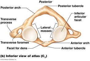
The atlas has no body or spinous process. It consists of anterior and posterior arches and two lateral masses. The superior surfaces of the lateral masses articulate with the occipital condyles, allowing the head to nod "yes."
Axis (C2)
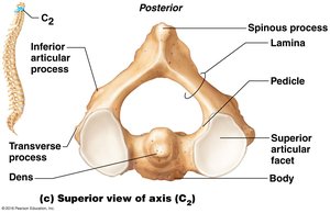
The axis has a body and processes like other vertebrae, but its major feature is the dens, a pivot for rotation of the atlas, allowing the head to rotate "no."
Muscle Attachments
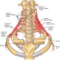
Scalenus muscles of the neck attach to the transverse processes of cervical vertebrae (C2–C7), elevating the first and second ribs.
Thoracic Vertebrae (T1–T12)
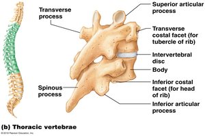
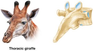
Thoracic vertebrae increase in size and articulate with ribs. Unique features include:
Heart-shaped body with demifacets for rib articulation
Circular vertebral foramen
Long, sharp spinous process pointing inferiorly
Transverse costal facets for rib articulation (except T11, T12)
Articular facets allow rotation
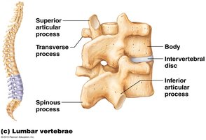
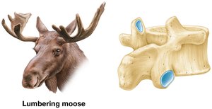
Lumbar Vertebrae (L1–L5)
Lumbar vertebrae are located in the lower back and receive the most stress. They have:
Massive, kidney-shaped bodies
Short, thick pedicles and laminae
Flat, hatchet-shaped spinous processes
Triangular vertebral foramen
Articular facets lock vertebrae together to prevent rotation
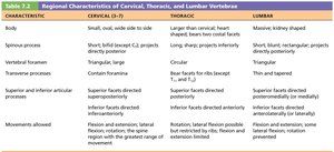
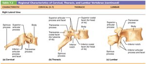
Comparison Table: Cervical, Thoracic, and Lumbar Vertebrae
Characteristic | Cervical (C3–C7) | Thoracic | Lumbar |
|---|---|---|---|
Body | Small, oval, side to side | Larger, heart-shaped | Massive, kidney-shaped |
Spinous process | Short, bifid (except C7) | Long, sharp, projects inferiorly | Short, flat, rectangular |
Vertebral foramen | Triangular, large | Circular | Triangular |
Transverse process | Contains foramen | Costal facets for ribs | No foramen |
Superior & inferior articular processes | Superior facets directed superoposteriorly; inferior facets directed inferoanteriorly | Superior facets directed posteriorly; inferior facets directed anteriorly | Superior facets directed posteromedially; inferior facets directed anterolaterally |
Movements allowed | Flexion, extension, lateral flexion, rotation | Rotation, limited flexion/extension | Flexion, extension, lateral flexion; rotation prevented |
Sacrum and Coccyx
Sacrum
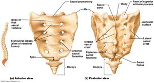
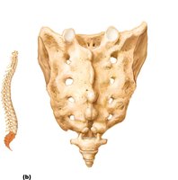
The sacrum is a triangular bone forming the posterior wall of the pelvis, made from five fused vertebrae (S1–S5). It articulates with L5, the coccyx, and the hip bones via auricular surfaces, forming sacroiliac joints.
Sacral promontory: Anterosuperior margin
Transverse ridges: Lines of fusion
Anterior and posterior sacral foramina: Openings for nerves and vessels
Median and lateral sacral crests: Roughened areas for muscle attachment
Sacral canal and hiatus: Continuation and opening of vertebral canal
Coccyx
The coccyx, or tailbone, is formed from three to five fused vertebrae. It has little function and fuses by age 20–30.
Thoracic Cage
Structure and Functions
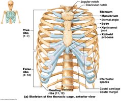
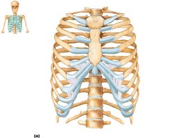
The thoracic cage is composed of thoracic vertebrae posteriorly, sternum and costal cartilages anteriorly, and ribs laterally. It protects vital organs, supports the shoulder girdles and upper limbs, and provides attachment sites for muscles.
Sternum
The sternum, or breastbone, consists of three fused bones:
Manubrium: Superior portion, articulates with clavicular notches and ribs 1 and 2
Body: Mid-portion, articulates with costal cartilages of ribs 2–7
Xiphoid process: Inferior end, site of muscle attachment; not ossified until ~age 40
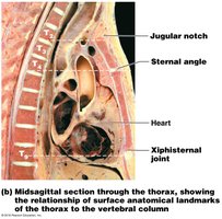
Important landmarks include the jugular notch, sternal angle, and xiphisternal joint.
Ribs
There are 12 pairs of ribs forming the sides of the thoracic cage:
True ribs (1–7): Attach directly to sternum via costal cartilages
False ribs (8–10): Attach indirectly by joining costal cartilage of rib above
Floating ribs (11–12): No attachment to sternum
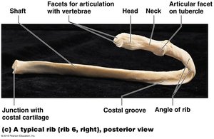
Main Parts of a Rib
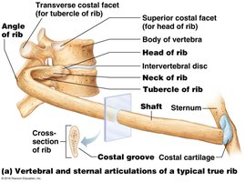
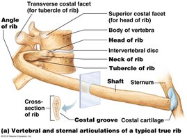
Shaft: Flat bone making up most of the rib
Costal groove: Houses nerves and vessels
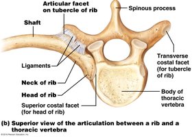
Head: Articulates with facets on bodies of two adjacent vertebrae
Neck: Constricted portion beyond head
Tubercle: Knoblike structure lateral to neck; articulates with transverse costal facet of same-numbered thoracic vertebra
Summary Table: Axial Skeleton Components
Region | Number of Bones | Main Function |
|---|---|---|
Cervical | 7 | Supports head, allows movement |
Thoracic | 12 | Articulates with ribs, protects organs |
Lumbar | 5 | Supports weight, resists stress |
Sacrum | 1 (5 fused) | Pelvic stability |
Coccyx | 1 (3–5 fused) | Minimal function |
Thoracic Cage | 25 (12 pairs ribs, sternum) | Protects organs, supports limbs |
Additional info: The notes have been expanded with academic context, definitions, and examples to ensure completeness and clarity for college-level Anatomy & Physiology students.
