 Indietro
The Brain and Cranial Nerves: Structure, Protection, and Cerebrospinal Fluid
Guida di studio - Note intelligenti
 Appunti personalizzati basati sui tuoi materiali, ampliati con definizioni chiave, esempi e contesto.
The Brain: Major Regions and Functions
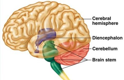
Overview of Brain Regions
The human brain is divided into four major regions, each with distinct anatomical features and specialized functions. Understanding these regions is fundamental to the study of neuroanatomy and physiology.
Cerebrum: Responsible for higher-order functions such as thought, memory, emotion, speech, and voluntary movement. It consists of two hemispheres and four lobes.
Cerebellum: Coordinates body movements, controls posture and balance, and is involved in motor learning and fine motor movement.
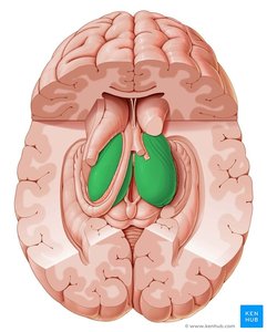
Diencephalon: Contains the thalamus and hypothalamus, serving as a relay station for sensory and motor signals and integrating nervous and endocrine functions.
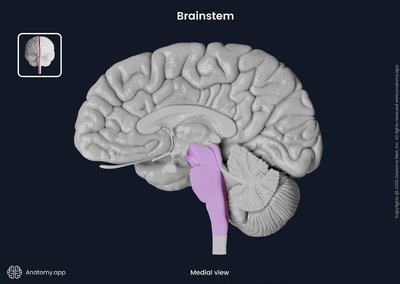
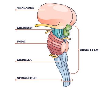
Brainstem: Processes and relays information between the spinal cord and higher brain regions. Includes the midbrain, pons, and medulla oblongata.
Cerebrum: Structure and Lobes
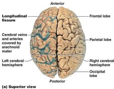
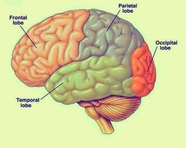
The cerebrum is the largest part of the brain, divided into left and right hemispheres and four lobes, each associated with specific functions.
Frontal lobe: Controls voluntary movement, reasoning, planning, and problem-solving.
Parietal lobe: Processes sensory information such as touch, temperature, and pain.
Temporal lobe: Responsible for auditory processing and memory.
Occipital lobe: Dedicated to visual processing.
Cerebellum
The cerebellum, often referred to as the "little brain," is located posterior to the cerebrum and is essential for coordination and balance.
Coordinates voluntary movements such as posture, balance, and fine motor skills.
Involved in motor learning and adaptation.
Diencephalon: Thalamus and Hypothalamus
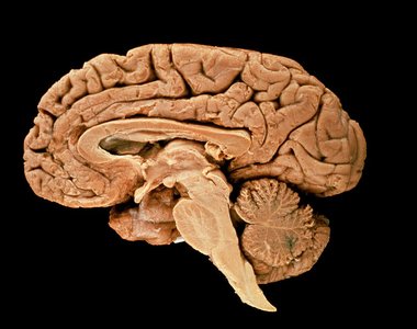
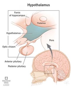
The diencephalon is situated deep within the brain and includes the thalamus and hypothalamus.
Thalamus: Acts as a relay station for sensory and motor signals to the cerebral cortex.
Hypothalamus: Integrates nervous and endocrine systems, regulates visceral functions, and connects to the pituitary gland.
Brainstem: Components and Functions
The brainstem is the conduit for information between the spinal cord and higher brain regions. It is composed of three main parts:
Midbrain: Controls visual and auditory reflexes.
Pons: Regulates breathing and communication between different parts of the brain.
Medulla oblongata: Controls autonomic functions such as heart rate and respiration.
Ventricular System of the Brain
Structure and Function
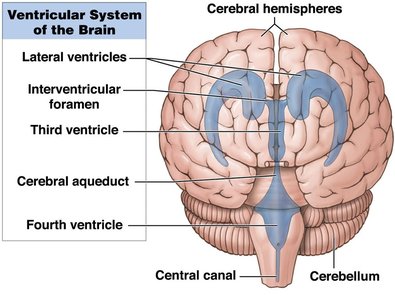
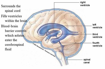
The ventricular system is a network of four fluid-filled cavities within the brain that produce and circulate cerebrospinal fluid (CSF), providing protection and support for the central nervous system.
Lateral ventricles: Located in each cerebral hemisphere.
Third ventricle: Found in the diencephalon.
Fourth ventricle: Extends into the medulla oblongata and connects to the central canal of the spinal cord.
Interventricular foramen: Connects lateral ventricles to the third ventricle.
Brain Protection and Support
Protective Layers
The brain is protected by several layers and structures that provide mechanical and physiological support.
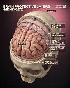
Skull (Cranium): Provides a rigid protective barrier against external trauma.
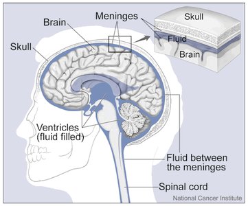
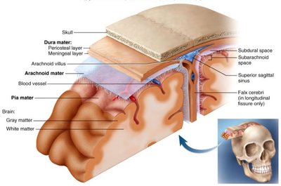
Meninges: Three membranes (dura mater, arachnoid mater, pia mater) that wrap the brain and spinal cord.
Cerebrospinal Fluid (CSF): Cushions the brain, reduces its effective weight, and transports nutrients and waste.
Blood-Brain Barrier: Selectively restricts passage of substances from the blood into the brain tissue.
Meninges: Structure and Function
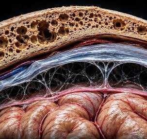
The meninges are three distinct layers that protect the brain and spinal cord:
Dura Mater: Tough, fibrous outer layer attached to the skull.
Arachnoid Mater: Web-like middle layer forming the subarachnoid space, which contains CSF.
Pia Mater: Delicate inner layer closely adhering to the brain surface and supporting blood vessels.
Dural Folds and Sinuses
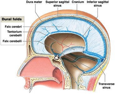
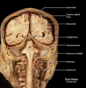
Dural folds are extensions of the dura mater that compartmentalize and stabilize the brain. Dural sinuses are venous channels that drain blood and CSF from the brain.
Falx cerebri: Separates the two cerebral hemispheres.
Tentorium cerebelli: Separates the cerebrum from the cerebellum.
Falx cerebelli: Separates the two hemispheres of the cerebellum.
Dural sinuses: Collect deoxygenated blood and CSF for removal via the internal jugular veins.
Arachnoid Villi and Granulations
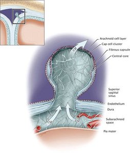
Arachnoid villi and granulations are structures that allow CSF to flow from the subarachnoid space into the dural sinuses, facilitating its removal from the brain.
Arachnoid villi: Act as one-way valves for CSF drainage.
Arachnoid granulations: Aggregations of villi, most commonly found along the superior sagittal sinus.
Cerebrospinal Fluid (CSF): Formation, Circulation, and Function
Formation of CSF
CSF is produced by the choroid plexus, a vascular tissue found in all four ventricles of the brain. Ependymal cells surrounding the capillaries secrete CSF and regulate its composition.
Choroid plexus: Produces CSF and forms the blood-CSF barrier.
Ependymal cells: Secrete CSF and remove wastes.
Circulation of CSF
CSF circulates through the ventricles, central canal, and subarachnoid space, providing cushioning and nutrient transport.
CSF flows from the lateral ventricles through the interventricular foramen to the third ventricle, then through the cerebral aqueduct to the fourth ventricle, and finally into the subarachnoid space.
CSF is absorbed into the bloodstream via arachnoid granulations.
Functions of CSF
Mechanical protection: Allows the brain to float, reducing its effective weight and cushioning neural tissue.
Chemical stability: Transports nutrients, chemical messengers, and wastes.
Found in: Lateral, third, and fourth ventricles, as well as the subarachnoid space.
Blood-Brain Barrier (BBB)
Structure and Function
The blood-brain barrier is a highly selective barrier that isolates the central nervous system from general circulation, protecting the brain from harmful substances.
Formed by: Tight junctions between capillary endothelial cells, astrocytes, and pericytes.
Selective permeability: Only lipid-soluble compounds (e.g., steroids, prostaglandins, alcohols, anesthetics) and gases (O2, CO2) can diffuse freely.
Astrocytes: Regulate BBB permeability by releasing chemicals.
Blood Supply to the Brain
Arteries: Internal carotid arteries and vertebral arteries deliver nutrients and oxygen.
Veins: Internal jugular veins remove blood via dural venous sinuses.
Summary Table: Major Brain Regions and Functions
Region | Main Function |
|---|---|
Cerebrum | Thought, memory, emotion, voluntary movement |
Cerebellum | Coordination, balance, motor learning |
Diencephalon | Sensory/motor relay, endocrine integration |
Brainstem | Autonomic functions, information relay |
Summary Table: Ventricular System
Ventricle | Location | Function |
|---|---|---|
Lateral | Cerebral hemispheres | CSF production and circulation |
Third | Diencephalon | CSF relay |
Fourth | Medulla oblongata | CSF relay to spinal cord |
Key Equations
CSF Flow Rate:
Blood-Brain Barrier Permeability:
Study Guide Questions
Name the major brain regions and functions of each region.
What are the four ventricles of the brain and where are they located?
Name the cranial meninges and describe their location and function.
What is the function of dural folds? Which layer forms dural folds and name the three dural folds.
What is the function of dural sinuses?
What is the function of arachnoid villi/granulations?
Which structure and glial cell type produces CSF? What are the cells involved?
Describe the CSF-blood barrier.
Functions and circulation of CSF (from ventricles across apertures into subarachnoid space).
Which glial cell makes up the blood-brain barrier?
What is the function of the blood-brain barrier? Explain the selective permeability of the BBB.
What are the arteries that supply blood to the brain and veins that carry blood away from the brain?
Additional info: Academic context was added to clarify the functions of each brain region, the structure and function of the ventricular system, and the mechanisms of brain protection and support. Tables and equations were inferred for completeness and exam preparation.
