 Indietro
The Brain and Cranial Nerves: Structure, Development, and Protection
Guida di studio - Note intelligenti
 Appunti personalizzati basati sui tuoi materiali, ampliati con definizioni chiave, esempi e contesto.
The Brain: Major Regions and Functions
Overview of Brain Regions
The adult human brain is the central organ of the nervous system, containing nearly 97% of the body's nervous tissue. It is divided into several major regions, each with distinct functions and anatomical features.
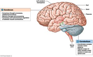
Cerebrum: The largest part, responsible for higher mental functions such as conscious thought, intellect, and memory. It is divided into left and right hemispheres.
Cerebellum: Coordinates repetitive body movements and maintains balance and posture.
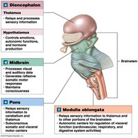
Diencephalon: Contains the thalamus (sensory relay and processing) and hypothalamus (emotions, autonomic functions, hormone production).
Brainstem: Relays information between the spinal cord and higher brain regions; includes the midbrain, pons, and medulla oblongata.
Surface Features of the Cerebrum
Gyri: Rounded elevations that increase surface area.
Sulci: Shallow depressions separating gyri.
Fissures: Deep grooves dividing major brain regions.
Development of the Brain
Embryonic Brain Vesicles
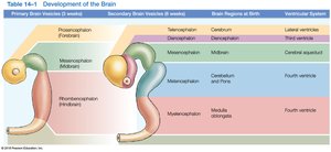
During embryonic development, the brain forms from the neural tube, which expands into three primary brain vesicles:
Prosencephalon (Forebrain)
Mesencephalon (Midbrain)
Rhombencephalon (Hindbrain)
These further differentiate into secondary vesicles, which give rise to the major adult brain regions and ventricles.
Ventricular System of the Brain
Structure and Function
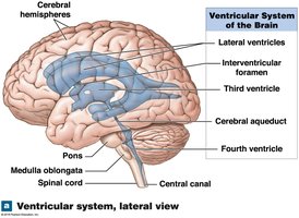
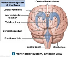
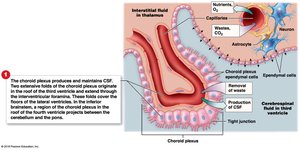
The brain contains a series of interconnected chambers called ventricles, filled with cerebrospinal fluid (CSF) and lined by ependymal cells. The main ventricles are:
Lateral ventricles: One in each cerebral hemisphere, separated by the septum pellucidum.
Third ventricle: Located in the diencephalon, connected to lateral ventricles via the interventricular foramen.
Fourth ventricle: Extends into the medulla oblongata, connected to the third ventricle by the cerebral aqueduct.
Protection and Support of the Brain
Cranial Meninges
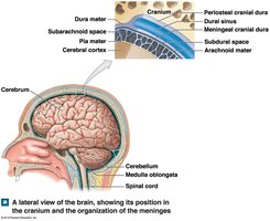
The brain is protected by three connective tissue layers called meninges:
Dura mater: Tough, outermost layer with periosteal and meningeal components.
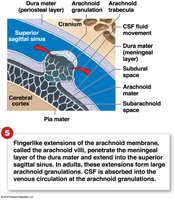
Arachnoid mater: Middle layer, separated from dura by the subdural space; subarachnoid space lies beneath, filled with CSF.
Pia mater: Delicate, innermost layer attached to the brain surface by astrocytes.
Dural Folds and Sinuses
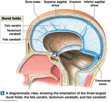
Dural folds are extensions of the dura mater that stabilize the brain and contain venous sinuses:
Falx cerebri: Between cerebral hemispheres.
Tentorium cerebelli: Separates cerebrum from cerebellum.
Falx cerebelli: Divides cerebellar hemispheres.
Cerebrospinal Fluid (CSF)
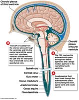
CSF surrounds all exposed surfaces of the CNS, providing support, cushioning, and transport of nutrients and wastes. It is produced by the choroid plexus in the ventricles and circulates through the ventricular system and subarachnoid space.
Brainstem: Medulla Oblongata, Pons, and Midbrain
Medulla Oblongata
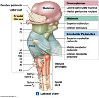
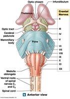
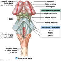
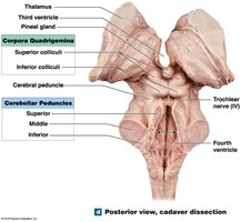
The medulla oblongata is the most inferior part of the brainstem, connecting the brain to the spinal cord. It contains nuclei that regulate autonomic functions (heart rate, blood pressure, digestion), relay sensory and motor information, and coordinate reflexes.
Pons
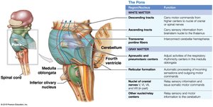
The pons contains sensory and motor nuclei for cranial nerves, nuclei involved in respiration, and tracts that relay information to and from the cerebellum.
Midbrain
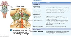
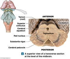
The midbrain processes visual and auditory information, generates reflexive motor responses, and maintains consciousness. Key structures include the tectum (corpora quadrigemina), tegmentum (red nucleus, substantia nigra), and cerebral peduncles.
Cerebellum: Structure and Function
Anatomy of the Cerebellum
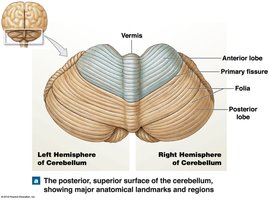
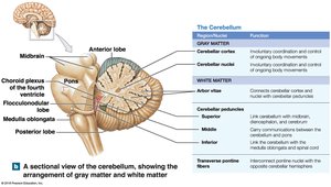
The cerebellum is responsible for adjusting postural muscles and fine-tuning conscious and subconscious movements. It consists of two hemispheres, the vermis, anterior and posterior lobes, and the flocculonodular lobe. The cerebellar cortex is highly folded, and the arbor vitae is the internal white matter.
Diencephalon: Thalamus and Hypothalamus
Thalamus
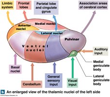
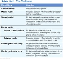
The thalamus acts as a relay and processing center for sensory information, filtering and transmitting signals to the cerebral cortex. It consists of several nuclei, each with specific functions related to sensory and motor integration.
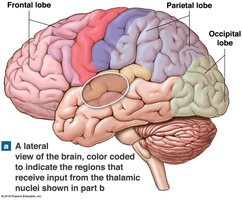
Nucleus/Body | Function |
|---|---|
Anterior nuclei | Part of the limbic system |
Medial nuclei | Integrate sensory information for projection to the frontal lobes |
Ventral nuclei | Project sensory information to the primary sensory cortex; relay information from cerebellum and basal nuclei to motor area of cerebral cortex |
Dorsal nuclei | Project information to parietal, occipitoparietal, and temporal cortex; may play a role in memory |
Pulvinar nuclei | Integrate sensory information for projection to association areas of cerebral cortex |
Lateral geniculate body | Projects visual information to the visual cortex; integrates sensory information and influences emotional states |
Medial geniculate body | Projects auditory information to the auditory cortex; integrates sensory information and influences emotional states |
Hypothalamus
Controls autonomic functions, emotions, and hormone production.
Regulates body temperature, circadian rhythms, hunger, thirst, and behavioral drives.
Connects to the pituitary gland via the infundibulum.
Summary
This guide covers the major anatomical regions of the brain, their development, protective structures, and key functions. Understanding these foundational concepts is essential for further study of the nervous system and its physiological roles.
