 Indietro
The Cardiovascular System: Structure, Function, and Blood Flow
Guida di studio - Note intelligenti
 Appunti personalizzati basati sui tuoi materiali, ampliati con definizioni chiave, esempi e contesto.
Cardiovascular System Overview
Introduction to the Cardiovascular System
The cardiovascular system is responsible for the transport of blood, nutrients, gases, and wastes throughout the body. It consists of the heart, blood vessels (arteries, veins, capillaries), and blood. The heart acts as a pump, circulating blood through two main circuits: the pulmonary and systemic circuits.
Heart: Pumps blood throughout the body.
Arteries: Carry blood away from the heart.
Veins: Carry blood toward the heart.
Blood Circulation Pathways
Pulmonary and Systemic Circuits
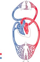
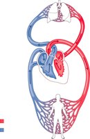
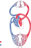
Blood circulates through two primary circuits: the pulmonary circuit and the systemic circuit. The pulmonary circuit transports blood between the heart and lungs for gas exchange, while the systemic circuit delivers oxygenated blood to the rest of the body.
Pulmonary Circuit: Right ventricle → pulmonary arteries → lungs (gas exchange) → pulmonary veins → left atrium.
Systemic Circuit: Left ventricle → aorta → body tissues (delivers oxygen) → veins → right atrium.
Heart Anatomy and Chambers
Location and Structure of the Heart
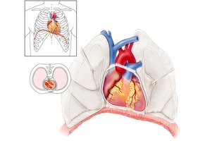
The heart is a muscular organ located slightly left of the midline, above the diaphragm, and is about the size of a fist. It is enclosed in a protective sac called the pericardium.
Pericardium: Double-walled sac that protects and anchors the heart.
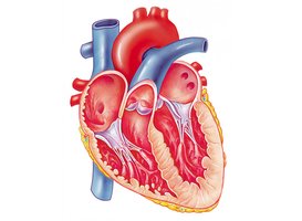
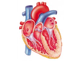
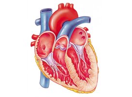
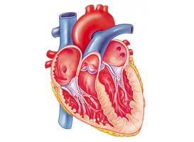
Chambers: Four chambers—right atrium, right ventricle, left atrium, left ventricle.
Layers of the Heart Wall
The heart wall consists of three layers: the epicardium (outer), myocardium (middle, muscle), and endocardium (inner lining).
Epicardium: Also called the visceral layer of serous pericardium.
Myocardium: Cardiac muscle tissue responsible for contraction.
Endocardium: Simple squamous epithelium lining the chambers.
Heart Valves and Blood Flow
Valves of the Heart
The heart contains four main valves that ensure unidirectional blood flow:
Atrioventricular (AV) valves: Tricuspid (right), Bicuspid/Mitral (left).
Semilunar valves: Pulmonary (right), Aortic (left).
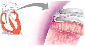
Chordae tendineae: Prevent valve inversion during contraction.
Blood Flow Through the Heart
Blood flows through the heart in a specific sequence, passing through each chamber and valve:
Deoxygenated blood enters the right atrium via the superior and inferior vena cava.
Passes through the tricuspid valve into the right ventricle.
Pumped through the pulmonary semilunar valve into the pulmonary trunk and arteries to the lungs.
Oxygenated blood returns via pulmonary veins to the left atrium.
Passes through the bicuspid (mitral) valve into the left ventricle.
Pumped through the aortic semilunar valve into the aorta and systemic circulation.
Heart Defects
Transposition of the Great Arteries
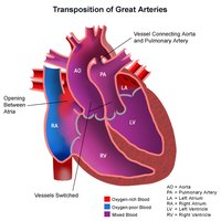
In this congenital defect, the positions of the aorta and pulmonary trunk are switched, resulting in improper circulation of oxygenated and deoxygenated blood. This can be life-threatening without surgical correction.
Microscopic Anatomy of Cardiac Muscle
Cardiac Muscle Structure
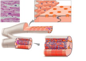
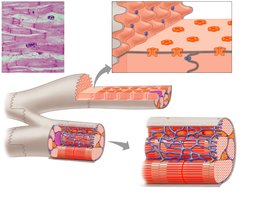
Cardiac muscle cells are striated and connected by specialized junctions called intercalated discs, which contain gap junctions and fascia adherens for coordinated contraction and strength.
Gap junctions: Allow rapid communication between cells.
Fascia adherens: Provide mechanical strength.
Conducting System of the Heart
Electrical Conduction Pathway
The heart's rhythmic contractions are coordinated by a specialized conduction system:
Sinoatrial (SA) node: Pacemaker, initiates impulse.
Atrioventricular (AV) node: Delays impulse before passing to ventricles.
AV bundle (Bundle of His), bundle branches, and Purkinje fibers: Distribute impulse through ventricles.
Blood Vessels: Structure and Function
General Structure of Blood Vessels
Blood vessels are composed of three layers (tunics):
Tunica intima: Endothelium (simple squamous epithelium) and subendothelial connective tissue.
Tunica media: Smooth muscle (controls vessel diameter).
Tunica externa: Connective tissue for support and protection.
Types of Blood Vessels
Arteries: Thick-walled, high pressure, carry blood away from heart.
Veins: Thinner walls, lower pressure, carry blood toward heart, contain valves.
Capillaries: Thin-walled, site of exchange between blood and tissues.
Capillary Types and Beds
Types of Capillaries
Continuous capillaries: Most common, tight junctions, found in muscle, skin, CNS.
Fenestrated capillaries: Pores for increased permeability, found in kidneys, intestines.
Sinusoidal capillaries: Large gaps, incomplete basement membrane, found in liver, spleen, bone marrow.
Blood: Composition and Function
Components of Blood
Blood is a connective tissue composed of plasma and formed elements:
Plasma: Liquid matrix, ~55% of blood, contains water, proteins (albumin, fibrinogen), nutrients, hormones, wastes.
Formed elements: Erythrocytes (RBCs), leukocytes (WBCs), platelets.
Erythrocytes (Red Blood Cells)
Function: Transport oxygen and carbon dioxide.
Structure: Biconcave, anucleate, filled with hemoglobin.
Lifespan: 3-4 months; produced in red bone marrow (hematopoiesis).
Leukocytes (White Blood Cells) and Platelets
Leukocytes: Immune response, can leave blood vessels (diapedesis).
Platelets: Cell fragments, essential for blood clotting.
Blood Groups and Transfusion
ABO and Rh Blood Groups
Blood types are determined by the presence of specific antigens (A, B, Rh) on erythrocyte membranes. The immune system produces antibodies against foreign antigens, which is critical for safe blood transfusions.
Type A: A antigen, anti-B antibody.
Type B: B antigen, anti-A antibody.
Type AB: A and B antigens, no anti-A or anti-B antibodies (universal recipient).
Type O: No A or B antigens, both anti-A and anti-B antibodies (universal donor).
Rh factor: Presence (+) or absence (−) of Rh antigen; Rh antibodies can cross the placenta.
Clinical Correlations
Anemia
Anemia is a condition characterized by a lower than normal number of red blood cells or hemoglobin, resulting in reduced oxygen delivery to tissues. Causes include iron deficiency, vitamin deficiencies, or chronic diseases.
Atherosclerosis and Myocardial Infarction
Atherosclerosis is the buildup of plaques in arterial walls, leading to narrowing and reduced blood flow. If blood supply to cardiac muscle is blocked, it can result in a myocardial infarction (heart attack).
Heart Murmurs and Valve Disorders
Abnormal heart sounds (murmurs) can result from valve defects such as mitral valve prolapse or stenosis, leading to inefficient blood flow and potential complications.
