 Indietro
The Cardiovascular System: The Heart – Structured Study Notes
Guida di studio - Note intelligenti
 Appunti personalizzati basati sui tuoi materiali, ampliati con definizioni chiave, esempi e contesto.
The Cardiovascular System: The Heart
Anatomy and Orientation of the Heart
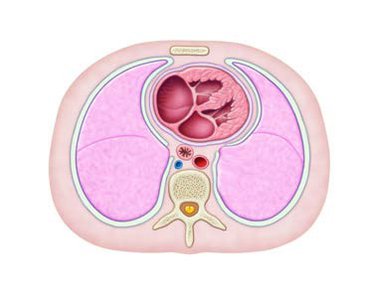
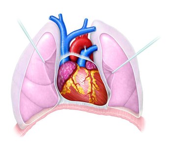
The heart is a muscular organ located in the mediastinum of the thoracic cavity, slightly left of the midsternal line. It is about the size of a fist and weighs between 250–350 grams. The heart is surrounded by the lungs laterally, the sternum anteriorly, and the vertebral column posteriorly. Its apex points inferiorly toward the left hip, and its base is directed toward the right shoulder.
Location: The heart is enclosed within the mediastinum, resting on the diaphragm.
Orientation: Two-thirds of its mass lies to the left of the midsternal line.
Function: Pumps blood through pulmonary and systemic circuits.
Pulmonary and Systemic Circuits
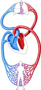
The heart functions as two pumps: the right side pumps blood through the pulmonary circuit (to the lungs), and the left side pumps blood through the systemic circuit (to the rest of the body). This ensures oxygen-poor blood is reoxygenated and oxygen-rich blood is distributed to tissues.
Pulmonary Circuit: Right ventricle → pulmonary arteries → lungs → pulmonary veins → left atrium.
Systemic Circuit: Left ventricle → aorta → body tissues → venae cavae → right atrium.
Coverings and Layers of the Heart Wall
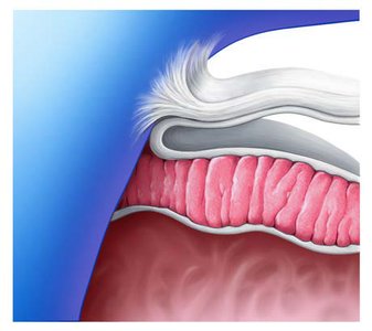
The heart is protected by a double-walled sac called the pericardium. The heart wall itself consists of three layers: epicardium, myocardium, and endocardium.
Fibrous Pericardium: Tough, dense connective tissue; protects and anchors the heart.
Serous Pericardium: Two layers (parietal and visceral/epicardium) with a lubricating pericardial cavity between them.
Epicardium: Visceral layer of serous pericardium; often infiltrated with fat.
Myocardium: Cardiac muscle; responsible for contraction.
Endocardium: Endothelial lining; continuous with blood vessels.
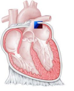
Cardiac Muscle Structure
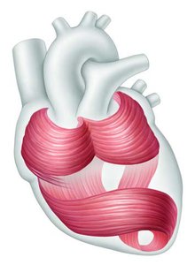
The myocardium is composed of spiral and circular bundles of cardiac muscle fibers, which are interconnected by intercalated discs containing desmosomes and gap junctions. This arrangement allows the heart to function as a coordinated unit (functional syncytium).
Intercalated Discs: Specialized junctions for mechanical and electrical coupling.
Cardiac Skeleton: Dense network of connective tissue reinforcing the myocardium.
Chambers of the Heart
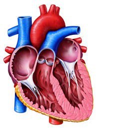
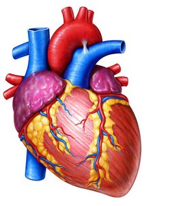
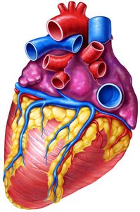
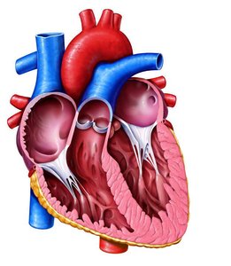
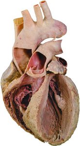
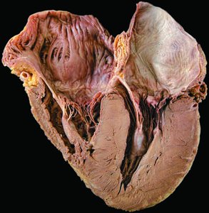
The heart has four chambers: two atria (receiving chambers) and two ventricles (pumping chambers). The right atrium receives oxygen-poor blood from the body, and the left atrium receives oxygen-rich blood from the lungs. The right ventricle pumps blood to the lungs, and the left ventricle pumps blood to the body.
Atria: Right atrium (systemic circuit), left atrium (pulmonary circuit).
Ventricles: Right ventricle (pulmonary trunk), left ventricle (aorta).
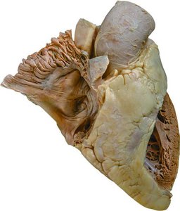
Internal Features: Interatrial and interventricular septa, pectinate muscles, crista terminalis, fossa ovalis, trabeculae carneae, papillary muscles.
Heart Valves
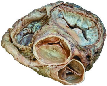
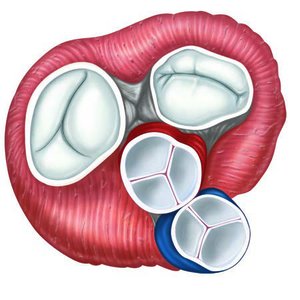
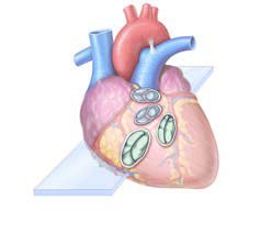
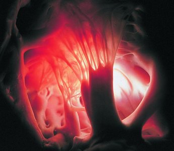
Valves ensure unidirectional blood flow through the heart. There are two sets: atrioventricular (AV) valves and semilunar (SL) valves.
AV Valves: Tricuspid (right), mitral/bicuspid (left); prevent backflow into atria.
SL Valves: Pulmonary (right), aortic (left); prevent backflow into ventricles.
Chordae Tendineae: Collagen cords anchoring AV valve flaps to papillary muscles.
Blood Flow Through the Heart
Blood flows from atria to ventricles, then to the lungs or body. The right side pumps to the pulmonary circuit, and the left side pumps to the systemic circuit. Equal volumes are pumped, but the left ventricle works harder due to higher resistance.
Pathway: Right atrium → right ventricle → pulmonary trunk → lungs → left atrium → left ventricle → aorta → body.
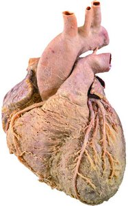
Coronary Circulation: Supplies the myocardium via coronary arteries and veins.
Chamber | Associated Vessel | Function |
|---|---|---|
Right Atrium | Superior/Inferior Vena Cava, Coronary Sinus | Receives oxygen-poor blood |
Right Ventricle | Pulmonary Trunk | Pumps blood to lungs |
Left Atrium | Pulmonary Veins | Receives oxygen-rich blood |
Left Ventricle | Aorta | Pumps blood to body |
Microscopic Anatomy and Physiology of Cardiac Muscle
Cardiac muscle cells are striated, short, branched, and interconnected. They contain abundant mitochondria and rely on aerobic respiration. Intercalated discs allow for coordinated contraction.
Desmosomes: Prevent cell separation during contraction.
Gap Junctions: Allow ion flow for electrical coupling.
Excitation-Contraction Coupling: Ca2+ influx triggers contraction.
Feature | Skeletal Muscle | Cardiac Muscle |
|---|---|---|
Structure | Long, cylindrical, multinucleate | Short, branched, 1-2 nuclei |
Gap Junctions | No | Yes |
Contraction | Individual motor units | As a unit (syncytium) |
ATP Supply | Aerobic/anaerobic | Aerobic only |
Electrical Events: Pacemaker Cells and Conduction System
The heart's rhythm is set by pacemaker cells in the sinoatrial (SA) node. The intrinsic conduction system coordinates depolarization and contraction.
SA Node: Pacemaker; initiates impulses.
AV Node: Delays impulse for atrial contraction.
AV Bundle, Bundle Branches, Purkinje Fibers: Conduct impulses to ventricles.
Autonomic Regulation of Heart Rate
The autonomic nervous system modulates heart rate and contractility. Sympathetic stimulation increases heart rate and force; parasympathetic stimulation decreases heart rate.
Sympathetic: Norepinephrine increases rate and contractility.
Parasympathetic: Acetylcholine decreases rate.
Electrocardiography (ECG)
An ECG records the electrical activity of the heart. The main waves are:
P Wave: Atrial depolarization.
QRS Complex: Ventricular depolarization.
T Wave: Ventricular repolarization.
The Cardiac Cycle: Mechanical Events
The cardiac cycle consists of all events during one heartbeat: atrial systole and diastole, followed by ventricular systole and diastole. Blood flow is governed by pressure changes and valve operation.
Phases: Ventricular filling, isovolumetric contraction, ventricular ejection, isovolumetric relaxation.
Heart Sounds: Lub (AV valves close), Dup (SL valves close).
Regulation of Cardiac Output
Cardiac output (CO) is the volume of blood pumped by each ventricle per minute. It is determined by heart rate (HR) and stroke volume (SV):
Formula:
Stroke Volume:
Factors: Preload (venous return), contractility, afterload (arterial pressure).
Factor | Effect on SV/CO |
|---|---|
Preload | Increases SV |
Contractility | Increases SV |
Afterload | Decreases SV |
Developmental Aspects of the Heart
The heart develops from mesoderm as a simple tube, which forms chambers and begins pumping blood by day 22 of gestation. Fetal circulation includes shunts (foramen ovale, ductus arteriosus) to bypass the lungs.
Congenital Defects: Septal defects, coarctation of the aorta, tetralogy of Fallot.
Age-related Changes: Valve sclerosis, reduced cardiac reserve, fibrosis, atherosclerosis.
Summary Table: Key Differences Between Skeletal and Cardiac Muscle
Feature | Skeletal Muscle | Cardiac Muscle |
|---|---|---|
Structure | Striated, long, cylindrical, multinucleate | Striated, short, branched, 1-2 nuclei |
Gap Junctions | No | Yes |
Contraction | Individual motor units | As a unit (syncytium) |
ATP Supply | Aerobic/anaerobic | Aerobic only |
Pacemaker Cells | No | Yes |
Clinical Connections
Pericarditis: Inflammation of the pericardium, can lead to cardiac tamponade.
Valve Disorders: Incompetent or stenotic valves increase cardiac workload.
Arrhythmias: Irregular heart rhythms, fibrillation, heart block.
Congestive Heart Failure: Inefficient pumping, can cause pulmonary or peripheral congestion.
Congenital Defects: Most common birth defects, can affect oxygenation or increase workload.
Key Equations
Review Questions and Practice
Describe the pathway of blood through the heart.
Explain the function and operation of heart valves.
Compare cardiac and skeletal muscle structure and function.
Interpret a normal ECG tracing.
Discuss the regulation of cardiac output.
Additional info: Academic context was added to clarify the physiological mechanisms, clinical relevance, and developmental aspects of the heart, as well as to provide self-contained explanations suitable for exam preparation.
