 Indietro
The Central Nervous System: Structure and Function
Guida di studio - Note intelligenti
 Appunti personalizzati basati sui tuoi materiali, ampliati con definizioni chiave, esempi e contesto.
The Central Nervous System
Brain Regions & Organization
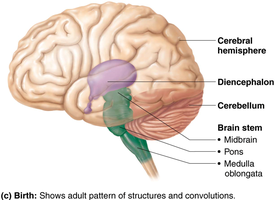
The adult human brain is divided into four main regions, each with distinct functions and anatomical features. Understanding these regions is essential for grasping the complexity of the central nervous system (CNS).
Cerebral Hemispheres: The largest part of the brain, responsible for higher cognitive functions.
Diencephalon: Central region containing key relay and regulatory structures.
Brain Stem: Includes the midbrain, pons, and medulla oblongata; controls vital functions.
Cerebellum: Coordinates movement and balance.
Folding During Development and CNS Structure
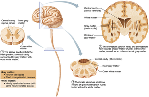
The CNS exhibits a basic structural pattern: a central cavity surrounded by gray matter, with white matter external to it. This arrangement is seen in both the brain and spinal cord, and is crucial for neural processing and communication.
Gray Matter: Composed of neuron cell bodies and short, nonmyelinated neurons.
White Matter: Contains myelinated and some nonmyelinated axons, facilitating rapid signal transmission.
Developmental Folding: Increases surface area and complexity, allowing for advanced functions.
Ventricles of the Brain
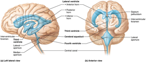
The brain contains fluid-filled chambers called ventricles, which are continuous with each other and the central canal of the spinal cord. These ventricles are filled with cerebrospinal fluid (CSF) and lined by ependymal cells, which are a type of neuroglial cell.
Lateral Ventricles: Paired, C-shaped chambers deep within each hemisphere.
Third Ventricle: Located in the diencephalon.
Fourth Ventricle: Located between the brainstem and cerebellum.
CSF Function: Cushions the brain, removes waste, and maintains chemical stability.
Ependymal Cells and Ventricular Lining
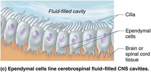
Ependymal cells line the ventricles and central canal, playing a critical role in the production and circulation of cerebrospinal fluid. Their cilia help move CSF throughout the CNS.
Cilia: Facilitate movement of CSF.
Barrier Function: Separate brain tissue from CSF-filled cavities.
The Cerebral Hemispheres
Surface Markings and Lobes
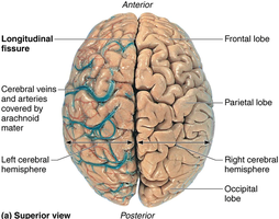
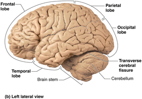
The cerebral hemispheres account for the majority of brain mass and are characterized by distinct surface features and divisions. These features are important for identifying functional regions.
Gyri: Ridges on the brain surface.
Sulci: Shallow grooves separating gyri.
Fissures: Deep grooves dividing major brain regions.
Longitudinal Fissure: Separates the two hemispheres.
Transverse Cerebral Fissure: Separates cerebrum from cerebellum.
Lobes of the Cerebral Hemispheres
Each hemisphere is divided into five lobes, named for the overlying skull bones. These lobes are responsible for different functions and are separated by sulci and fissures.
Frontal Lobe: Associated with reasoning, planning, movement, and problem-solving.
Parietal Lobe: Processes sensory information related to touch, pressure, and spatial orientation.
Temporal Lobe: Involved in hearing, memory, and speech.
Occipital Lobe: Responsible for visual processing.
Insula: Buried under portions of the other lobes; involved in taste, visceral sensation, and autonomic control.
Regions of the Cerebral Hemispheres
Each hemisphere contains three basic regions:
Cerebral Cortex: Superficial layer of gray matter, responsible for conscious thought and awareness.
White Matter: Internal region, composed of myelinated axons.
Basal Nuclei: Deep within the white matter, involved in motor control and coordination.
Cerebral Cortex: Functional Areas
The cerebral cortex is the "executive suite" of the brain, responsible for processing sensory information, initiating voluntary movement, and supporting communication, memory, learning, and understanding. It contains three types of functional areas:
Motor Areas: Control voluntary movement.
Sensory Areas: Provide conscious awareness of sensation.
Association Areas: Integrate information, support memory, language, planning, reasoning, and decision-making.
Contralateral Control and Lateralization
Each hemisphere of the cerebral cortex controls and receives input from the opposite side of the body (contralateral control). Additionally, certain functions are lateralized, meaning they are specialized to one hemisphere:
Left Hemisphere: Language, logic, mathematics.
Right Hemisphere: Emotional expression, creativity, spatial awareness.
Integrated Cortical Function
Conscious behavior involves multiple regions of the cortex working together. Sensory, motor, and association areas interact continuously to produce thought and behavior.
Motor and Sensory Areas
Motor Areas
Located in the frontal lobe, motor areas control and direct voluntary movements. The primary motor cortex, found in the precentral gyrus, initiates skeletal muscle contractions. The motor homunculus is a map showing the cortical area devoted to each body region, with larger areas for regions requiring fine motor control.
Sensory Areas
Sensory areas of the cerebral cortex are responsible for the conscious awareness and interpretation of sensory information. The primary somatosensory cortex, located in the postcentral gyrus of the parietal lobe, receives input from the skin, muscles, joints, and tendons, allowing spatial discrimination and mapping sensory input (somatosensory homunculus).
The Diencephalon
Thalamus, Hypothalamus, and Epithalamus
The diencephalon is the central region of the brain, consisting of three paired gray-matter structures: thalamus, hypothalamus, and epithalamus. All three enclose the third ventricle.
Thalamus: The largest part, acts as a relay station for sensory information (except smell).
Hypothalamus: Major homeostatic control center, regulates autonomic functions and endocrine activity.
Epithalamus: Contains the pineal gland, which secretes melatonin to regulate sleep-wake cycles.
The Brain Stem
Midbrain, Pons, and Medulla Oblongata
The brainstem connects the spinal cord to the cerebrum and controls automatic behaviors necessary for survival. It consists of three regions:
Midbrain: Contains important relay and reflex centers.
Pons: Bridges higher brain centers and the spinal cord; relays impulses between motor cortex and cerebellum.
Medulla Oblongata: Autonomic reflex center; regulates cardiovascular, respiratory, and other vital functions.
The Cerebellum
Coordination and Balance
The cerebellum processes input from the cortex, brain stem, and sensory receptors to provide precise, coordinated movements of skeletal muscles. It also plays a major role in maintaining balance.
Protection of the Brain
Meninges
The brain is protected by three layers of connective tissue called meninges:
Dura Mater: Tough, outermost layer; consists of periosteal and meningeal layers.
Arachnoid Mater: Middle layer with spiderweb-like extensions; contains CSF and blood vessels.
Pia Mater: Delicate, innermost layer; clings tightly to the brain and contains blood vessels.
Cerebrospinal Fluid (CSF)
CSF forms a liquid cushion around the brain, providing buoyancy, protection, and nourishment. It is produced by the choroid plexus and maintained by ependymal cells.
Blood Brain Barrier (BBB)
The BBB maintains a stable environment for the brain by restricting the passage of substances from the blood. It consists of tight junctions between endothelial cells, a thick basement membrane, and specific transport mechanisms.
The Spinal Cord
Reflex Center and Conduction Pathway
The spinal cord is enclosed in the vertebral column and protected by bone, meninges, and CSF. It provides two-way communication between the brain and body and serves as a major reflex center.
Epidural Space: Cushion of fat and veins between vertebrae and dura mater.
CSF: Fills the subarachnoid space between arachnoid and pia maters.
Site of Lumbar Puncture: Dural and arachnoid membranes extend to the sacrum, allowing safe access for CSF sampling.
Additional info: The notes above expand on the original content by providing definitions, examples, and context for each anatomical structure and function, ensuring completeness and academic quality for exam preparation.
