 Indietro
The Digestive System: Functional Anatomy, Physiology, and Regulation
Guida di studio - Note intelligenti
 Appunti personalizzati basati sui tuoi materiali, ampliati con definizioni chiave, esempi e contesto.
The Digestive System
Overview and Functions
The digestive system is responsible for breaking down food, absorbing nutrients, and eliminating waste. It consists of a series of organs and tissues that work together to provide fuel for the body and maintain homeostasis.
Ingestion: Intake of food into the oral cavity.
Mechanical digestion: Physical breakdown of food by chewing and mixing.
Chemical digestion: Enzymatic breakdown of macromolecules.
Secretion: Release of digestive enzymes, acids, and buffers.
Absorption: Uptake of nutrients into the bloodstream or lymphatic system.
Defecation: Elimination of indigestible material.
Defense: Mucosa-associated lymphoid tissue (MALT) provides immune protection.
Major Organs and Accessory Structures
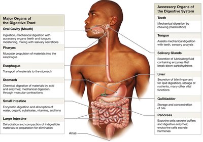
The digestive tract is composed of the oral cavity, pharynx, esophagus, stomach, small intestine, and large intestine. Accessory organs include the pancreas, gallbladder, liver, salivary glands, tongue, and teeth.
Oral Cavity: Begins digestion; senses, analyzes, and lubricates food.
Pharynx: Passageway for food, liquids, and air; initiates swallowing.
Esophagus: Muscular tube transporting food to the stomach.
Stomach: Stores and digests food; produces intrinsic factor and forms chyme.
Small Intestine: Main site for enzymatic digestion and absorption.
Large Intestine: Reabsorbs water, compacts feces, absorbs vitamins.
Accessory Organs: Salivary glands (secrete enzymes), liver (produces bile), gallbladder (stores bile), pancreas (secretes enzymes and hormones).
Histology and Functional Anatomy
Layers of the Digestive Tract
The digestive tract has four major layers, each with specialized functions:
Mucosa: Inner lining; contains mucous membrane, circular folds, villi, and glands.
Submucosa: Dense connective tissue; contains blood vessels, lymphatics, and the submucosal plexus.
Muscular Layer: Smooth muscle; responsible for peristalsis and segmentation, controlled by the myenteric plexus.
Serosa: Outer membrane; present in organs within the peritoneal cavity.
Movement of Food
Peristalsis: Rhythmic contractions move the bolus along the tract.
Segmentation: Churning and mixing of contents, mainly in the small intestine.
Oral Cavity, Pharynx, and Esophagus
Oral Cavity
The oral cavity is lined by mucosa and contains teeth, tongue, and salivary glands.
Teeth: Perform mastication (chewing).
Tongue: Muscular organ; compresses and distorts food, secretes lingual lipase.
Salivary Glands: Three pairs; secrete saliva containing water, mucins, ions, buffers, antibodies, and enzymes.
Pharynx and Esophagus
Pharynx: Common passageway; muscles initiate swallowing (deglutition).
Esophagus: Muscular tube; upper third contains skeletal muscle, lower third smooth muscle; lower esophageal sphincter prevents backflow.
Swallowing Phases
Buccal Phase: Voluntary; tongue moves bolus, soft palate closes nasopharynx.
Pharyngeal Phase: Involuntary; epiglottis closes, larynx elevates, breathing stops, bolus moves to esophagus.
Esophageal Phase: Involuntary; peristalsis moves bolus to stomach.
Stomach
Structure and Function
The stomach is a muscular organ with three layers (longitudinal, circular, oblique) and prominent folds (rugae) for expansion.
Functions: Storage, mechanical digestion, chemical digestion, intrinsic factor production.
Chyme: Mixture of food and gastric secretions.
Stomach Lining and Secretory Cells
Parietal Cells: Secrete intrinsic factor (for vitamin B12 absorption) and hydrochloric acid (HCl).
Chief Cells: Secrete pepsinogen, converted to pepsin by HCl; pepsin digests proteins.
G Cells: Secrete gastrin, stimulating parietal and chief cells and stomach contractions.
Small Intestine
Structure and Regions
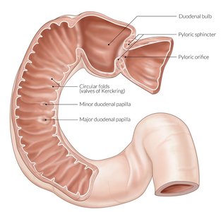
The small intestine is divided into the duodenum, jejunum, and ileum. It is the primary site for digestion and absorption.
Duodenum: Receives chyme and enzymes from pancreas and liver.
Jejunum: Main site for chemical digestion and nutrient absorption.
Ileum: Controls flow into the large intestine.
Histology and Absorption
Villi: Finger-like projections increasing surface area; contain capillaries and lacteals.
Brush Border: Microvilli on epithelial cells; site of brush border enzymes.
Lacteal: Lymphatic capillary for absorption of lipids.
Submucosal Glands: Produce alkaline mucus to buffer acids.
Intestinal Hormones
Gastrin: Increases stomach motility and enzyme secretion.
Secretin: Stimulates bile and pancreatic enzyme secretion; released when duodenal pH falls.
Cholecystokinin (CCK): Stimulates digestive enzyme production and bile ejection; released when chyme enters duodenum.
Gastric Inhibitory Peptide (GIP): Inhibits gastric activity; released when fats and glucose enter intestine.
Regulation of Digestive Activity
Neural and Hormonal Control
Digestive activities are regulated by the central nervous system (CNS), enteric nervous system (ENS), and hormones.
Submucosal and Myenteric Plexuses: Components of the ENS; control local reflexes and muscle contractions.
Cephalic Phase: CNS prepares stomach for food.
Gastric Phase: Local reflexes triggered by stretch and chemoreceptors.
Intestinal Phase: Controls gastric emptying; stimulates intestinal hormones.
Pancreas, Liver, and Gallbladder
Pancreas
Exocrine Cells: Produce digestive enzymes (carbohydrases, lipases, nucleases, proteases).
Enzymes: Trypsin, chymotrypsin, carboxypeptidase; released into duodenum.
Liver
Metabolic Regulation: Adjusts circulating nutrients, stores excess.
Hematological Regulation: Processes blood, removes toxins.
Bile Production: Bile emulsifies lipids for digestion.
Liver Lobules: Hepatocytes surrounded by sinusoids; portal triad (portal vein, hepatic artery, bile duct).
Gallbladder
Function: Stores and concentrates bile; releases bile upon CCK stimulation.
Large Intestine
Structure and Function
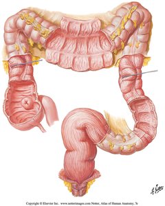
The large intestine receives material from the small intestine and is responsible for water reabsorption, compaction of feces, vitamin absorption, and storage of waste.
Cecum: Initial segment; slow movement of material.
Colon: Reabsorbs water, bile salts, vitamins; processes waste.
Rectum: Storage of feces; initiates defecation reflex.
Defecation
Defecation Reflex: Triggered by stretch receptors in rectum; involves voluntary relaxation of external anal sphincter and involuntary relaxation of internal sphincter.
Hemorrhoids: Caused by excessive straining and distension of rectal veins.
Summary Table: Digestive System Organs and Functions
Organ | Main Function |
|---|---|
Oral Cavity | Mechanical and chemical digestion, sensory analysis |
Pharynx | Initiates swallowing |
Esophagus | Transports food to stomach |
Stomach | Mechanical and chemical digestion, intrinsic factor production |
Small Intestine | Enzymatic digestion, absorption of nutrients |
Large Intestine | Water reabsorption, feces compaction, vitamin absorption |
Liver | Bile production, metabolic regulation |
Gallbladder | Bile storage and concentration |
Pancreas | Enzyme and buffer secretion |
Key Terms and Concepts
Peristalsis: Rhythmic contraction of smooth muscle to move food.
Segmentation: Mixing of intestinal contents.
Intrinsic Factor: Protein required for vitamin B12 absorption.
Chyme: Semi-fluid mixture of food and gastric secretions.
Brush Border Enzymes: Enzymes on microvilli that complete digestion.
Emulsification: Breakdown of fat droplets by bile salts.
Sample Questions for Review
What are the main functions of the digestive system?
Which cells secrete pepsinogen in the stomach?
What is the role of brush border enzymes?
Which hormone stimulates bile release from the gallbladder?
Why does the liver have dual circulation?
Equations and Formulas
General formula for carbohydrate digestion:
Protein digestion:
Lipid digestion:
Additional info: The notes have been expanded to include definitions, examples, and academic context for clarity and completeness.
