 Indietro
The Digestive System: Structure, Function, and Regulation
Guida di studio - Note intelligenti
 Appunti personalizzati basati sui tuoi materiali, ampliati con definizioni chiave, esempi e contesto.
The Digestive System Overview
Alimentary Canal and Accessory Organs
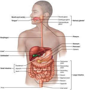
The digestive system consists of the alimentary canal (gastrointestinal tract) and accessory organs. The alimentary canal is a continuous muscular tube extending from the mouth to the anus, responsible for digesting food and absorbing nutrients. Accessory organs, such as the teeth, tongue, gallbladder, salivary glands, liver, and pancreas, aid in digestion by producing secretions or facilitating mechanical breakdown.
Alimentary canal organs: Mouth, pharynx, esophagus, stomach, small intestine, large intestine, anus
Accessory organs: Teeth, tongue, gallbladder, salivary glands, liver, pancreas
Digestive Processes
Steps in Food Processing
Food is processed through six main steps in the digestive system, each involving specific organs and mechanisms:
Ingestion: Intake of food into the mouth
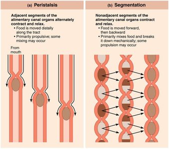
Propulsion: Movement of food through the alimentary canal, including swallowing (voluntary) and peristalsis (involuntary)
Mechanical breakdown: Physical breakdown of food by chewing, mixing with saliva, churning in the stomach, and segmentation in the intestines
Digestion: Enzymatic breakdown of complex food molecules into absorbable units
Absorption: Passage of digested nutrients from the GI tract into blood or lymph
Defecation: Elimination of indigestible substances as feces
Overview of Digestive Functions by Organ
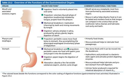
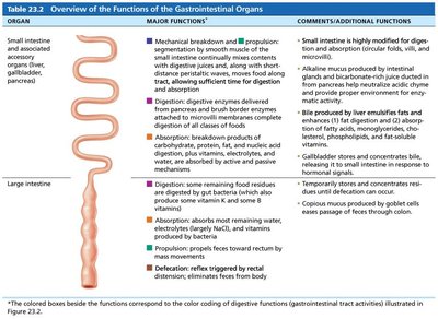
The digestive organs perform distinct functions, including mechanical and chemical digestion, absorption, and propulsion. The following tables summarize these functions:
Organ | Major Functions | Comments/Additional Functions |
|---|---|---|
Mouth and associated accessory organs | Ingestion, propulsion, mechanical breakdown, digestion | Mouth serves as a receptacle; saliva cleanses and lubricates; enzymes begin starch digestion |
Pharynx and esophagus | Propulsion (swallowing and peristalsis) | Primarily food chutes; mucus produced helps lubricate food passage |
Stomach | Mechanical breakdown, propulsion, digestion, absorption | Hydrochloric acid and pepsin digest proteins; mucus protects stomach lining; some absorption of water, alcohol, and drugs |
Organ | Major Functions | Comments/Additional Functions |
|---|---|---|
Small intestine and associated accessory organs | Mechanical breakdown, propulsion, digestion, absorption | Highly modified for digestion and absorption; bile emulsifies fats; enzymes digest nutrients; absorption of water, electrolytes, and nutrients |
Large intestine | Digestion, absorption, propulsion, defecation | Temporary storage and concentration of residues; absorption of water and electrolytes; formation and elimination of feces |
Peritoneum and Abdominal Cavity
Structure and Function of the Peritoneum
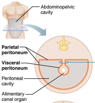
The peritoneum is a serous membrane lining the abdominal cavity, consisting of the visceral peritoneum (covering digestive organs) and parietal peritoneum (lining the body wall). The peritoneal cavity is the fluid-filled space between these layers, providing lubrication for organ movement.
Visceral peritoneum: Covers external surfaces of digestive organs
Parietal peritoneum: Lines the abdominal wall
Peritoneal cavity: Contains lubricating serous fluid
Histology of the Alimentary Canal
Layers of the Alimentary Canal
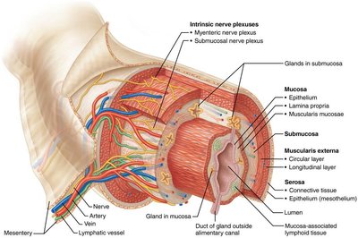
All digestive organs share four basic layers (tunics): mucosa, submucosa, muscularis externa, and serosa. Each layer has specialized functions and structures.
Mucosa: Lines the lumen; secretes mucus, enzymes, and hormones; absorbs nutrients; protects against infection
Submucosa: Contains blood and lymphatic vessels, nerve plexuses, and elastic tissue
Muscularis externa: Responsible for segmentation and peristalsis; contains circular and longitudinal muscle layers
Serosa: Outermost layer; visceral peritoneum
Enteric Nervous System
Intrinsic Nerve Plexuses and Regulation
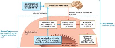
The GI tract has its own nervous system, the enteric nervous system (ENS), which regulates digestive activity through intrinsic nerve plexuses. The ENS controls motility, secretion, and blood flow, and interacts with the central nervous system via short and long reflexes.
Submucosal nerve plexus: Regulates glands and smooth muscle in mucosa
Myenteric nerve plexus: Controls GI tract motility
Short reflexes: Mediated by ENS; respond to local stimuli
Long reflexes: Involve CNS; respond to internal and external stimuli
Mouth and Associated Organs
Structure and Function
The mouth is the entry point for food, where mechanical and chemical digestion begins. Associated organs include the tongue, salivary glands, and teeth, each contributing to food processing and digestion.
Tongue: Grips, repositions, and mixes food; forms bolus; initiates swallowing, speech, and taste
Salivary glands: Produce saliva to cleanse mouth, dissolve food, moisten food, and begin starch digestion
Teeth: Responsible for mastication (chewing), breaking food into smaller fragments
Pharynx and Esophagus
Swallowing and Propulsion
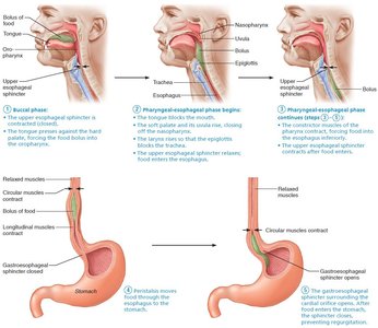
Food passes from the mouth into the pharynx and esophagus, which serve as conduits for food movement to the stomach. Swallowing (deglutition) involves coordinated muscle contractions in two phases: buccal (voluntary) and pharyngeal-esophageal (involuntary).
Buccal phase: Voluntary contraction of the tongue
Pharyngeal-esophageal phase: Involuntary, controlled by the swallowing center in the brainstem
Stomach
Structure and Function
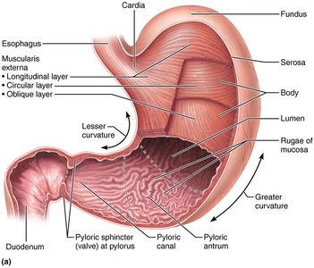
The stomach is a temporary storage organ that initiates protein digestion and converts food into chyme. It has specialized folds (rugae) and a modified muscularis externa with an additional oblique layer for churning and mixing food.
Rugae: Folds in the stomach mucosa that allow expansion
Muscularis externa: Includes circular, longitudinal, and oblique muscle layers
Microscopic Anatomy and Gland Cells
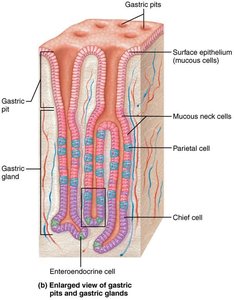
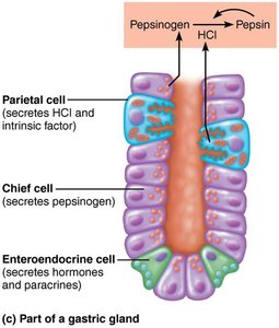
The stomach mucosa contains gastric pits leading to gastric glands, which produce gastric juice. Four main types of gland cells are present:
Mucous neck cells: Secrete thin, acidic mucus
Parietal cells: Secrete hydrochloric acid (HCl) and intrinsic factor
Chief cells: Secrete pepsinogen (inactive enzyme) and lipases
Enteroendocrine cells: Secrete hormones and paracrines (e.g., serotonin, histamine, somatostatin, gastrin)
Mucosal Barrier and Clinical Imbalances
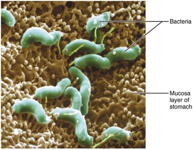
The stomach is protected by a mucosal barrier consisting of bicarbonate-rich mucus, tight junctions between epithelial cells, and rapid cell turnover. Disruption of this barrier can lead to gastritis and peptic ulcers, often caused by Helicobacter pylori or NSAIDs.
Regulation of Gastric Secretion
Phases of Gastric Secretion
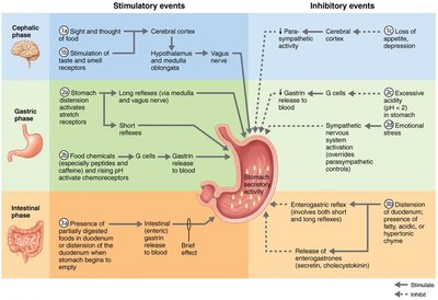
Gastric secretion is regulated by neural and hormonal mechanisms and occurs in three phases:
Cephalic phase: Triggered by sight, smell, taste, or thought of food
Gastric phase: Initiated by food entry into the stomach; involves stretch and chemical stimuli
Intestinal phase: Begins as chyme enters the small intestine; includes both stimulatory and inhibitory components
Hormones and Paracrines in Digestion
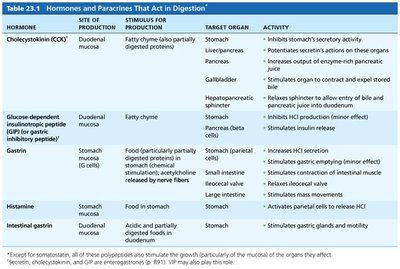
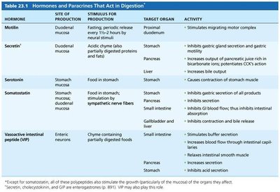
Several hormones and paracrines regulate digestive activity, including cholecystokinin (CCK), secretin, gastrin, histamine, serotonin, and somatostatin. These molecules influence secretion, motility, and absorption in various digestive organs.
Hormone | Site of Production | Stimulus | Target Organ | Activity |
|---|---|---|---|---|
Cholecystokinin (CCK) | Duodenal mucosa | Fatty chyme | Stomach, liver, pancreas, gallbladder | Inhibits stomach secretion, stimulates bile and pancreatic juice release |
Gastrin | Stomach G cells | Food, acetylcholine | Stomach, small intestine | Stimulates HCl secretion, increases motility |
Secretin | Duodenal mucosa | Acidic chyme | Stomach, pancreas, liver | Inhibits gastric secretion, stimulates bile and pancreatic juice |
Histamine | Stomach mucosa | Food in stomach | Stomach | Activates parietal cells to release HCl |
Summary
The digestive system is a complex network of organs and tissues that work together to process food, absorb nutrients, and eliminate waste. Understanding the structure, function, and regulation of each component is essential for mastering anatomy and physiology.
