 Indietro
The Endocrine System: Structure, Function, and Disorders
Guida di studio - Note intelligenti
 Appunti personalizzati basati sui tuoi materiali, ampliati con definizioni chiave, esempi e contesto.
The Endocrine System
Overview of Endocrine Organs
The endocrine system consists of glands that secrete hormones directly into the bloodstream, regulating a wide range of physiological processes. Unlike other systems, the endocrine system's organs do not work together for a single function but are grouped due to their common method of hormone secretion.
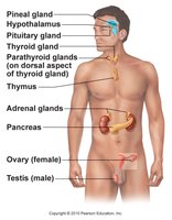
Endocrine glands include the pituitary, thyroid, parathyroid, adrenal, pancreas, gonads, pineal, and thymus.
Some organs, such as the heart and kidneys, also have endocrine functions.
Hormones are classified as amino acid-based or steroid-based, affecting their mechanism of action.
Hormone Action and Regulation
Hormone Mechanisms
Hormones exert their effects by binding to specific receptors on or within target cells. The presence and number of receptors determine tissue sensitivity to hormones.
Agonists: Molecules that mimic natural hormones and activate receptors.
Antagonists: Molecules that block hormone action by occupying receptors.
Physiological, pharmacological, and subphysiological levels: Refer to normal, abnormally high, and abnormally low hormone concentrations, respectively.
Up-regulation: Increase in receptor number, enhancing sensitivity.
Down-regulation: Decrease in receptor number, reducing sensitivity.
Second Messenger Systems
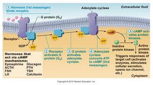
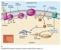
Amino acid-based hormones cannot cross cell membranes and act via second messengers such as cyclic AMP (cAMP) and calcium ions (Ca2+).
cAMP Pathway: Hormone binds receptor → G protein activation → Adenylate cyclase activation → ATP converted to cAMP → cAMP activates protein kinase A → Cellular response.
Calcium Pathway: Hormone binds receptor → G protein activation → Phospholipase activation → PIP2 split into DAG and IP3 → IP3 releases Ca2+ from ER → Ca2+ activates calmodulin → Cellular response.

Steroid Hormone Action
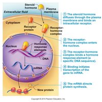
Steroid and thyroid hormones are lipid-soluble and diffuse across cell membranes to bind intracellular receptors, directly affecting gene transcription and protein synthesis.
Hormone enters cell → Binds cytoplasmic or nuclear receptor → Hormone-receptor complex binds DNA → Alters transcription → Changes protein synthesis → Cellular response.
Response is slower, often taking hours to days.
Major Endocrine Glands and Their Hormones
Pituitary Gland
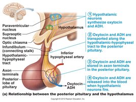
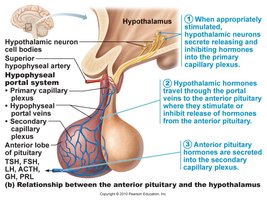
The pituitary gland, located at the base of the brain, consists of two distinct parts: the anterior (adenohypophysis) and posterior (neurohypophysis) pituitary. It is often called the 'Master Gland' due to its regulatory role over other endocrine glands.
Anterior Pituitary: Releases protein-based hormones via second messengers. Key hormones include:
Growth Hormone (GH, somatotropin): Stimulates tissue growth, especially muscles and bones.
Thyroid Stimulating Hormone (TSH, thyrotropin): Stimulates thyroid hormone production.
Adrenocorticotropic Hormone (ACTH, corticotropin): Stimulates adrenal cortex to release glucocorticoids.
Follicle Stimulating Hormone (FSH): Stimulates ovarian follicle growth and sperm production.
Luteinizing Hormone (LH): Triggers ovulation and testosterone production.
Prolactin: Stimulates milk production in females.
Posterior Pituitary: Stores and releases two hormones produced in the hypothalamus:
Antidiuretic Hormone (ADH, vasopressin): Promotes water retention by kidneys.
Oxytocin: Stimulates uterine and mammary duct contractions.
Adrenal Glands
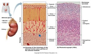
The adrenal glands, located above the kidneys, consist of an outer cortex and inner medulla, each producing distinct hormones.
Adrenal Medulla: Secretes catecholamines (epinephrine and norepinephrine) for the fight-or-flight response.
Adrenal Cortex: Produces three classes of steroid hormones:
Mineralocorticoids (e.g., aldosterone): Regulate sodium, potassium, and blood volume.
Glucocorticoids (e.g., cortisol): Affect metabolism and stress response.
Sex steroids (androgens): Supplement gonadal hormones.
Thyroid Gland
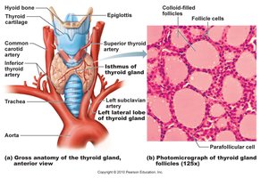
The thyroid gland, located below the larynx, produces hormones essential for metabolism and growth. It consists of follicles filled with colloid and surrounded by follicular and parafollicular cells.
T3 (triiodothyronine) and T4 (thyroxine): Regulate metabolic rate and development.
Calcitonin: Lowers blood calcium levels, especially important in youth.
Thyroid hormone synthesis involves iodide uptake, thyroglobulin formation, and hormone release upon TSH stimulation.

Parathyroid Glands
The parathyroid glands are four small glands on the posterior surface of the thyroid. They regulate blood calcium levels through secretion of parathyroid hormone (PTH).
PTH: Increases blood calcium by stimulating osteoclasts, enhancing kidney calcium retention, and promoting vitamin D activation for intestinal absorption.
Maintaining calcium homeostasis is critical for muscle and nerve function.

Pancreas
The pancreas has both endocrine and exocrine functions. The endocrine portion consists of the Islets of Langerhans, which regulate blood glucose levels.
Alpha cells: Secrete glucagon, raising blood glucose during fasting.
Beta cells: Secrete insulin, lowering blood glucose after meals.
Imbalance leads to diabetes mellitus, characterized by excessive urination, thirst, and hunger.
Type 1 diabetes: Insulin deficiency; Type 2 diabetes: Insulin resistance.


Pineal Gland
The pineal gland, located in the brain, secretes melatonin, which regulates sleep cycles. Melatonin secretion is inhibited by light and is highest at night.
Thymus
The thymus, located behind the sternum, is important for immune system maturation, especially T-cell development. It is largest in newborns and regresses after puberty.
Gonads
The gonads (testes and ovaries) produce sex hormones essential for reproductive function and secondary sexual characteristics.
Testes: Produce androgens (testosterone) for male development.
Ovaries: Produce estrogens and progesterone for female reproductive cycle.
Endocrine Disorders
Growth Hormone Disorders
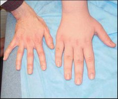
Gigantism: Excess GH in youth leads to abnormal growth and acromegaly in adults.
Dwarfism: GH deficiency results in short stature and underdeveloped features.




Diabetes Insipidus
Diabetes insipidus: ADH deficiency causes excessive urine production and dehydration.

Thyroid Disorders
Hyperthyroidism (Graves disease): Excess thyroid hormone causes goiter, tachycardia, nervousness, high metabolism, and exophthalmos (bulging eyes).
Hypothyroidism: Deficiency leads to fatigue, low metabolism, weight gain, and myxedema. In children, it causes cretinism (stunted growth and mental retardation).


Adrenal Disorders
Conn's Disease: Excess aldosterone causes high blood pressure and muscle weakness.
Cushing's Disease: Excess glucocorticoids cause moon face, buffalo hump, poor wound healing, and abnormal fat distribution.
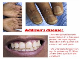
Addison's Disease: Deficiency of aldosterone and cortisol leads to low blood pressure, dehydration, and skin bronzing.

Summary Table: Major Endocrine Glands and Their Hormones
Gland | Main Hormones | Primary Functions |
|---|---|---|
Pituitary (anterior) | GH, TSH, ACTH, FSH, LH, PRL | Growth, metabolism, reproduction, lactation |
Pituitary (posterior) | ADH, Oxytocin | Water balance, uterine/mammary contraction |
Thyroid | T3, T4, Calcitonin | Metabolism, calcium regulation |
Parathyroid | PTH | Calcium homeostasis |
Adrenal (cortex) | Aldosterone, Cortisol, Androgens | Electrolyte balance, stress response, sex hormones |
Adrenal (medulla) | Epinephrine, Norepinephrine | Fight-or-flight response |
Pancreas | Insulin, Glucagon | Blood glucose regulation |
Pineal | Melatonin | Sleep regulation |
Thymus | Thymosin | Immune cell maturation |
Gonads | Testosterone, Estrogen, Progesterone | Reproduction, secondary sex characteristics |
Additional info: Academic context was added to clarify hormone mechanisms, disorders, and gland functions. All images included are directly relevant to the adjacent explanations.
