 Indietro
The Heart: Structure, Function, and Circulation
Guida di studio - Note intelligenti
 Appunti personalizzati basati sui tuoi materiali, ampliati con definizioni chiave, esempi e contesto.
The Heart: Structure, Function, and Circulation
Overview and Location of the Heart
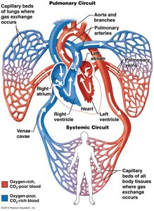
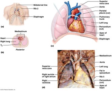
The heart is a muscular organ essential for pumping blood throughout the body via the pulmonary and systemic circuits. It is cone-shaped, weighs between 250-350 grams, and is located in the thoracic cavity, specifically within the mediastinum. The apex of the heart lies to the left of the midline, while the base is the broad posterior surface.
Thoracic cavity: The heart is the largest organ in the mediastinum.
Four corners: Anatomical landmarks include the costal cartilages of the second, third, fifth, and sixth ribs.
Coverings of the Heart
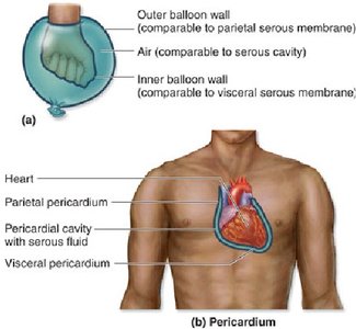
The heart is surrounded by the pericardium, which consists of two main sections: the fibrous pericardium and the serous pericardium. These layers protect the heart and reduce friction during contraction.
Fibrous pericardium: Dense irregular connective tissue that anchors the heart to the diaphragm and great vessels.
Serous pericardium: Composed of two layers:
Parietal layer: Lines the internal surface of the fibrous pericardium.
Visceral layer (epicardium): Directly covers the heart.
Pericardial cavity: Space between the parietal and visceral layers containing serous fluid for lubrication.
Layers of the Heart Wall
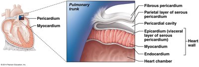
The heart wall is composed of three distinct layers, each with specialized structure and function.
Epicardium: The visceral layer of the serous pericardium, often mixed with fat.
Myocardium: The thickest layer, consisting of cardiac muscle tissue responsible for contraction and pumping blood. Muscle fibers are arranged in circular and spiral bundles.
Endocardium: A layer of simple squamous epithelium resting on thin connective tissue, lining the heart chambers and valves.
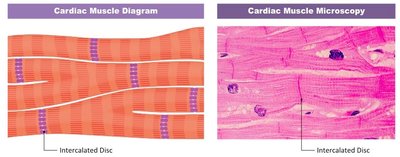
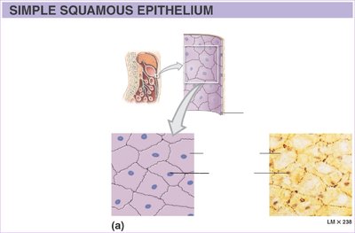
Internal and External Divisions of the Heart
The heart is divided into four chambers: two atria and two ventricles. Internal septa and external sulci mark the boundaries between these chambers.
Interatrial septum: Separates the right and left atria.
Interventricular septum: Separates the right and left ventricles.
Coronary sulcus: Horizontally separates atria from ventricles.
Anterior and posterior interventricular sulci: Mark the external boundaries of the ventricles.
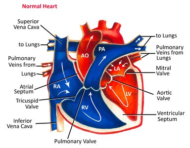
Chambers of the Heart and Their Structures
Each chamber of the heart has unique anatomical features and functions.
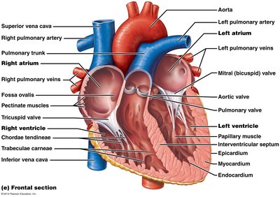
Right Atrium: Receives oxygen-poor blood from the superior and inferior vena cava and coronary sinus. Contains pectinate muscles and an auricle to increase capacity.
Right Ventricle: Receives blood from the right atrium via the tricuspid valve and pumps it into the pulmonary circuit through the pulmonary trunk. Internal features include trabeculae carneae, papillary muscles, and chordae tendineae.
Left Atrium: Receives oxygen-rich blood from the lungs via pulmonary veins. Blood passes to the left ventricle through the bicuspid (mitral) valve.
Left Ventricle: Forms the apex of the heart and pumps blood through the systemic circuit via the aortic semilunar valve. Internal features are similar to the right ventricle.
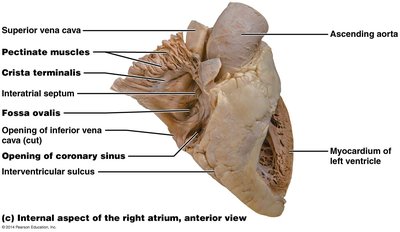
Major Arteries and Veins
The heart is connected to major arteries and veins that facilitate blood flow to and from the body and lungs.
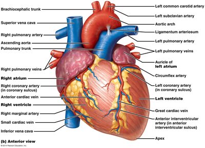
Arteries: Pulmonary trunk (from right ventricle), aorta (from left ventricle).
Veins: Superior and inferior vena cava (to right atrium), pulmonary veins (to left atrium).
Heart Valves: Types and Functions
The heart contains four valves that ensure unidirectional blood flow and prevent backflow.
Atrioventricular (AV) valves: Tricuspid (right) and bicuspid/mitral (left). Prevent backflow into atria during ventricular contraction. Supported by chordae tendineae and papillary muscles.
Semilunar valves: Pulmonary (right ventricle) and aortic (left ventricle). Prevent backflow into ventricles during relaxation.
Heart Sounds
Heart sounds are produced by the closure of valves during the cardiac cycle. "Lub" corresponds to AV valve closure, and "Dub" to semilunar valve closure. Abnormal sounds may indicate pathology.
Lub: Closure of AV valves.
Dub: Closure of semilunar valves.
Listening regions: Pulmonary, aortic, mitral, and tricuspid valve areas.
Pathway of Blood Through the Heart
Blood flows through the heart in a specific sequence, passing through both pulmonary and systemic circuits.
Oxygen-poor blood enters the right atrium via the superior and inferior vena cava.
Blood passes through the tricuspid valve into the right ventricle.
Right ventricle pumps blood through the pulmonary valve into the pulmonary trunk and arteries to the lungs.
Oxygen-rich blood returns to the left atrium via pulmonary veins.
Blood passes through the bicuspid (mitral) valve into the left ventricle.
Left ventricle pumps blood through the aortic valve into the aorta and systemic circulation.
Conducting System of the Heart
The heart's intrinsic conducting system coordinates rhythmic contractions. Specialized cardiac muscle cells generate and conduct impulses.
Sinoatrial (SA) node: Pacemaker, sets the rate of contraction.
Internodal pathway: Conducts impulses to the atrioventricular (AV) node.
AV node: Delays impulse before sending to AV bundle.
AV bundle: Connects atria to ventricles.
Bundle branches: Conduct impulses through interventricular septum.
Purkinje fibers: Stimulate contractile cells of ventricles.
Blood Supply to the Heart
The heart receives its own blood supply via the coronary arteries and returns deoxygenated blood through cardiac veins.
Coronary arteries: Right and left coronary arteries arise from the base of the aorta.
Left coronary artery: Branches into anterior interventricular artery (LAD) and circumflex artery.
Right coronary artery: Branches into marginal artery and posterior interventricular artery (PDA).
Cardiac veins: Great, middle, and small cardiac veins drain into the coronary sinus, which returns blood to the right atrium.
Disorders of the Heart
Several disorders can affect the heart's function and structure.
Coronary artery disease: Includes atherosclerosis, angina pectoris, myocardial infarction (heart attack), and silent ischemia.
Heart failure: Progressive weakening, inability to meet oxygen demands, congestive heart failure (CHF), and pulmonary arterial hypertension.
Arrhythmias: Abnormal heart rhythms, including atrial fibrillation (impulses circle within atrial myocardium, risk of clots and stroke) and ventricular fibrillation (rapid, random impulses, common cause of cardiac arrest).
Key Terms and Definitions
Epicardium: Outer layer of the heart wall, also known as visceral pericardium.
Myocardium: Middle, muscular layer responsible for contraction.
Endocardium: Inner layer lining chambers and valves.
Septum: Wall dividing chambers (interatrial and interventricular).
Sulcus: Groove marking external boundaries.
Valve: Structure ensuring unidirectional blood flow.
Chordae tendineae: Fibrous cords preventing AV valve inversion.
Purkinje fibers: Specialized conducting fibers in ventricles.
Summary Table: Heart Chambers and Associated Structures
Chamber | Main Function | Associated Structures |
|---|---|---|
Right Atrium | Receives oxygen-poor blood | Superior/inferior vena cava, coronary sinus, pectinate muscles, auricle |
Right Ventricle | Pumps blood to lungs | Tricuspid valve, pulmonary valve, trabeculae carneae, papillary muscles, chordae tendineae |
Left Atrium | Receives oxygen-rich blood | Pulmonary veins, bicuspid (mitral) valve, auricle |
Left Ventricle | Pumps blood to body | Aortic valve, trabeculae carneae, papillary muscles, chordae tendineae |
Summary Table: Heart Valves
Valve | Location | Function |
|---|---|---|
Tricuspid (AV) | Between right atrium and ventricle | Prevents backflow into right atrium |
Bicuspid (Mitral, AV) | Between left atrium and ventricle | Prevents backflow into left atrium |
Pulmonary (Semilunar) | Between right ventricle and pulmonary trunk | Prevents backflow into right ventricle |
Aortic (Semilunar) | Between left ventricle and aorta | Prevents backflow into left ventricle |
Key Equations
Cardiac Output: The amount of blood pumped by the heart per minute.
Stroke Volume: The amount of blood pumped by a ventricle in one contraction.
Additional info: Academic context was added to clarify anatomical terms, functions, and clinical relevance. Images were included only when directly relevant to the explanation of the paragraph.
