 Indietro
The Heart: Structure, Function, and Physiology
Guida di studio - Note intelligenti
 Appunti personalizzati basati sui tuoi materiali, ampliati con definizioni chiave, esempi e contesto.
The Heart: Structure, Function, and Physiology
Location and Orientation of the Heart
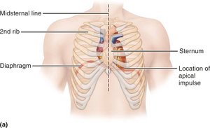
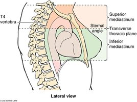
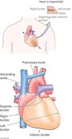
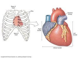
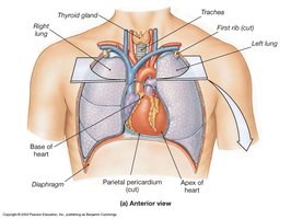
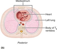
The heart is centrally located within the thoracic cavity, specifically in the mediastinum, posterior to the sternum and between the lungs. Its orientation is such that the base is superior and the apex points inferiorly and to the left.
Mediastinum: The central compartment of the thoracic cavity, containing the heart, great vessels, trachea, and esophagus.
Base: The broad superior portion where the great veins and arteries attach.
Apex: The pointed inferior tip, located near the left fifth intercostal space.
Borders: The heart presents superior, inferior, left, and right borders.
Coverings of the Heart: The Pericardium
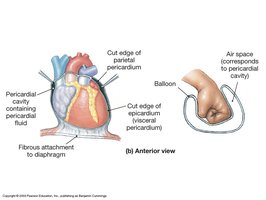
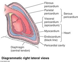
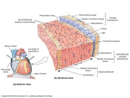
The heart is enclosed by the pericardium, a double-walled sac that provides protection, reduces friction, and anchors the heart within the thoracic cavity.
Parietal pericardium: Lines the inner surface of the pericardial sac.
Visceral pericardium (epicardium): Covers the outer surface of the heart.
Pericardial cavity: Space between the parietal and visceral layers, filled with pericardial fluid to reduce friction.
Structure of the Heart Wall
The heart wall consists of three distinct layers, each with specialized functions.
Epicardium: The outermost layer, also known as the visceral pericardium.
Myocardium: The thick, muscular middle layer responsible for contraction.
Endocardium: The innermost layer, lining the heart chambers and covering the valves.
Cardiac Muscle Tissue
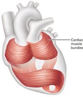
Cardiac muscle tissue forms the bulk of the myocardium and is specialized for continuous, rhythmic contraction.
Striated: Contains myofibrils similar to skeletal muscle.
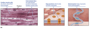
Intercalated discs: Specialized junctions that connect cardiac muscle cells, containing gap junctions and desmosomes.
Aerobic respiration: Cardiac cells are highly dependent on oxygen and contain numerous mitochondria.
Gap junctions: Allow electrical impulses to pass rapidly between cells, enabling coordinated contraction.
Desmosomes: Provide mechanical strength by binding cells together.
Differences Between Cardiac and Skeletal Muscle Tissue
Cardiac muscle differs from skeletal muscle in several key aspects, including cell structure, energy requirements, and contraction mechanisms.
Cell shape: Cardiac muscle cells are shorter and branched, while skeletal muscle cells are long and cylindrical.
Intercalated discs: Present only in cardiac muscle, facilitating rapid electrical communication.
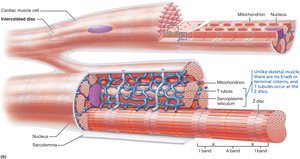
Mitochondria: Cardiac muscle cells contain more mitochondria, supporting their resistance to fatigue.
Sarcoplasmic reticulum: Less elaborate in cardiac muscle; no terminal cisterns or triads.
T tubules: Wider and less numerous in cardiac muscle, entering at Z discs.
Feature | Skeletal Muscle | Cardiac Muscle |
|---|---|---|
Structure | Striated, long, cylindrical, multinucleate | Striated, short, branched, one or two nuclei per cell |
Gap junctions | No | Yes |
Contracts as a unit | No | Yes |
T tubules | Abundant | Fewer |
Sarcoplasmic reticulum | Elaborate | Less elaborate |
Source of Ca2+ for contraction | Sarcoplasmic reticulum | Sarcoplasmic reticulum and extracellular fluid |
Pacemaker cells | No | Yes |
Tetanic contractions | Possible | No |
ATP supply | Aerobic and anaerobic | Aerobic only (more mitochondria) |
Superficial and Internal Anatomy of the Heart
The heart's external and internal features are essential for its function as a pump.
Auricle: Expandable extension of each atrium.
Coronary sulcus: Groove separating atria from ventricles.
Interatrial septum: Separates left and right atria.
Interventricular septum: Separates left and right ventricles.
Valves: Ensure one-way flow of blood between chambers.
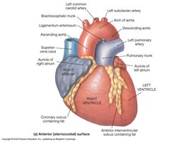
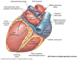
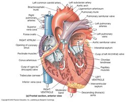
Chambers and Valves of the Heart
The heart contains four chambers and four main valves, each with specific roles in blood flow.
Right atrium: Receives deoxygenated blood from the systemic circuit.
Right ventricle: Pumps blood to the pulmonary circuit via the pulmonary trunk.
Left atrium: Receives oxygenated blood from the pulmonary veins.
Left ventricle: Pumps oxygenated blood to the systemic circuit via the aorta.
Valves: Tricuspid (right AV), bicuspid/mitral (left AV), pulmonary semilunar, and aortic semilunar valves.
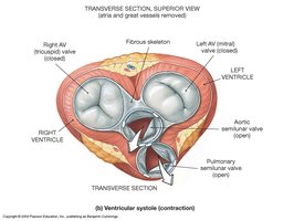
Fibrous Skeleton of the Heart
The fibrous skeleton is a network of connective tissue that provides structural support, anchors valves, and electrically isolates the atria from the ventricles.
Stabilizes contractile cells and valves.
Distributes force of contraction.
Adds strength and elasticity.
Physically isolates atria from ventricles.
Coronary Circulation
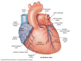
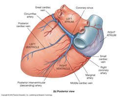
Coronary circulation supplies blood to the myocardium, ensuring the heart receives adequate oxygen and nutrients.
Coronary arteries: Originate at the base of the ascending aorta; right and left coronary arteries branch to supply the heart.
Coronary veins: Collect deoxygenated blood from the myocardium and return it to the right atrium via the coronary sinus.
Cardiac Conduction System
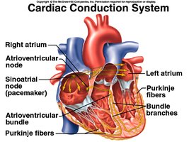
The cardiac conduction system coordinates the heartbeat through specialized pacemaker and conducting cells.
SA node: Pacemaker, initiates heartbeat.
AV node: Electrical gateway to ventricles.
AV bundle and bundle branches: Pathways for signals to descend to the apex.
Purkinje fibers: Spread signals throughout ventricular myocardium.
Electrical Activity of the Heart
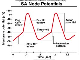
Electrical impulses originate in the SA node and propagate through the heart, resulting in coordinated contraction.
Pacemaker potential: Unstable resting membrane potential allows spontaneous depolarization.
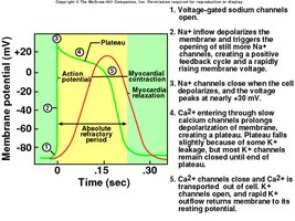
Action potential: Involves rapid influx of Ca2+ and efflux of K+.
Myocardial action potential: Longer duration than skeletal muscle, includes plateau phase.
Comparison of Cardiac and Skeletal Muscle Action Potentials
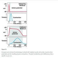
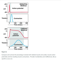
Cardiac muscle action potentials are longer and include a plateau phase, preventing tetanic contractions and ensuring efficient blood ejection.
Duration: Cardiac AP lasts 200+ ms; skeletal AP lasts 1–2 ms.
Plateau phase: Unique to cardiac muscle, prolongs contraction.
Refractory period: Prevents tetanus in cardiac muscle.
Feature | Skeletal Muscle | Cardiac Muscle |
|---|---|---|
Action potential duration | 1–2 ms | 200+ ms |
Contraction duration | 15–100 ms | 200+ ms |
Plateau phase | No | Yes |
Tetanic contractions | Possible | No |
Electrocardiogram (ECG)
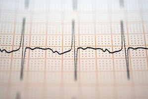
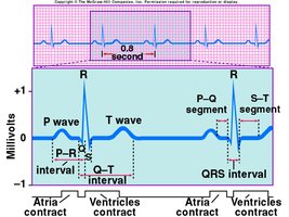
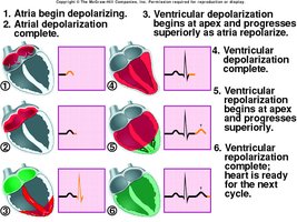
An ECG records the electrical activity of the heart, providing information about the timing and sequence of atrial and ventricular depolarization and repolarization.
P wave: Atrial depolarization.
QRS complex: Ventricular depolarization (atrial repolarization obscured).
T wave: Ventricular repolarization.
The Cardiac Cycle
The cardiac cycle consists of systole (contraction) and diastole (relaxation), with each cycle lasting about 0.8 seconds.
Systole: Contraction phase, blood is ejected from chambers.
Diastole: Relaxation phase, chambers fill with blood.
Heart rate: Approximately 75 beats per minute.
Blood Flow Through the Heart
Blood flows through the heart in a specific sequence, passing through chambers and valves to ensure efficient circulation.
Right atrium → right ventricle → pulmonary arteries → lungs → left atrium → left ventricle → aorta → body
Heart Valves and Their Function
Heart valves permit blood flow in only one direction, preventing regurgitation and creating characteristic heart sounds.
AV valves: Tricuspid and mitral, prevent backflow into atria.
Semilunar valves: Pulmonary and aortic, prevent backflow into ventricles.
Heart sounds: S1 (lubb) from AV valve closure, S2 (dupp) from semilunar valve closure.
Major Events of the Cardiac Cycle
The cardiac cycle includes ventricular filling, isovolumetric contraction, ventricular ejection, and isovolumetric relaxation.
Ventricular filling: Blood flows from atria to ventricles during diastole.
Isovolumetric contraction: Ventricles contract with all valves closed.
Ventricular ejection: Semilunar valves open, blood is ejected.
Isovolumetric relaxation: Ventricles relax, all valves closed.
Cardiac Output (CO)
Cardiac output is the volume of blood ejected by each ventricle per minute.
Formula:
Resting values: 70 ml/beat × 75 beats/min = 5,250 ml/min
Exercise: CO can increase to 21–35 L/min in athletes.
Stroke Volume (SV)
Stroke volume is the difference between end-diastolic volume (EDV) and end-systolic volume (ESV).
Formula:
Factors: Preload, contractility, afterload
Preload and Afterload
Preload is the stretch in ventricular myocardium before contraction; afterload is the pressure opposing valve opening.
Increased preload: Increases stroke volume.
Increased afterload: Decreases stroke volume.
Heart Rate and Its Regulation
Heart rate varies with age, sex, and autonomic nervous system activity.
Tachycardia: HR > 100 bpm
Bradycardia: HR < 60 bpm
Chronotropic agents: Influence HR; positive agents increase, negative agents decrease.
Autonomic control: Cardioacceleratory center (sympathetic) increases HR; cardioinhibitory center (parasympathetic) decreases HR.
Summary
The heart is a complex organ with specialized structures and functions that ensure efficient circulation. Understanding its anatomy, muscle tissue, conduction system, and physiological processes is essential for comprehending cardiovascular health and disease.
