 Indietro
The Human Skull: Cranial and Facial Bones
Guida di studio - Note intelligenti
 Appunti personalizzati basati sui tuoi materiali, ampliati con definizioni chiave, esempi e contesto.
The Human Skull
Overview of Skull Structure
The human skull is a complex structure composed of 22 bones, which account for about 8% of body weight. These bones are grouped into two main categories: the axial skeleton (skull, vertebral column, rib cage) and the appendicular skeleton (limbs and girdles). The skull itself is divided into cranial and facial bones, each serving distinct functions in protection, support, and sensory input.
Cranial bones: Form the protective case for the brain and house organs of hearing and balance.
Facial bones: Create the framework of the face, secure teeth, and anchor facial muscles.
Cranial Bones
Major Cranial Bones and Their Functions
The cranial bones are mostly flat and are united by immovable joints called sutures. There are eight cranial bones: paired parietal and temporal bones, and unpaired frontal, occipital, sphenoid, and ethmoid bones. These bones surround and protect the brain, and their internal surfaces are divided into three distinct regions called cranial fossae.
Frontal bone: Dome-shaped, forms the forehead and roof of the orbits; contributes to the anterior cranial fossa.
Parietal bones: Paired, form the superior and lateral aspects of the skull; bulk of the cranial vault.
Occipital bone: Single bone at the base of the skull; forms the posterior aspect and walls of the posterior cranial fossa.
Temporal bones: Paired, form the inferior and lateral aspects of the skull and parts of the cranial floor; contain regions such as squamous, tympanic, and petrous parts.
Sphenoid bone: Complex bone that articulates with all other cranial bones; forms the base of the middle cranial fossa.
Ethmoid bone: Cube-shaped, lies deep between the orbits and nasal cavities; forms the roof of the nasal cavity and part of the anterior cranial fossa.
Cranial Fossae
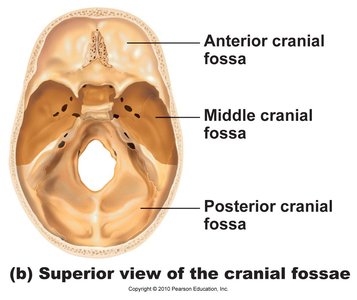
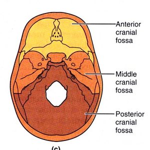
The cranial base is divided into three fossae: anterior, middle, and posterior. These depressions accommodate different parts of the brain.
Anterior cranial fossa: Houses the frontal lobes of the brain.
Middle cranial fossa: Contains the temporal lobes.
Posterior cranial fossa: Supports the cerebellum.
Sutures of the Skull
Sutures are immovable joints that firmly unite the bones of the adult skull. The main sutures include:
Coronal suture: Connects the frontal bone to the parietal bones.
Squamous suture: Connects the parietal bone to the temporal bone.
Lambdoid suture: Connects the occipital bone to the parietal bones.
Sagittal suture: Connects the two parietal bones.
Facial Bones
Major Facial Bones and Their Functions
The facial skeleton consists of 14 bones, of which only the mandible and vomer are unpaired. The paired bones include maxillae, zygomatic, nasal, lacrimal, palatine, and inferior nasal conchae. These bones form the framework of the face, house sensory organs, and provide openings for air and food passage.
Mandible: Lower jawbone; largest and strongest facial bone; holds lower teeth and forms the chin.
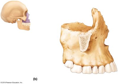
Maxillae: Fused medially; hold upper teeth and form the anterior portion of the hard palate.
Zygomatic bones: Cheekbones; articulate with maxilla, frontal, and temporal bones.
Nasal bones: Form the bridge of the nose.
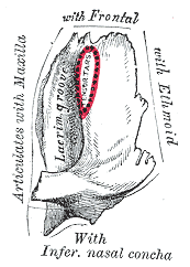
Lacrimal bones: Fingernail-shaped bones in the medial orbit; each has a lacrimal fossa for the lacrimal sac.
Palatine bones: L-shaped; form part of the hard palate and nasal cavity.
Vomer: Thin, single bone forming part of the nasal septum.
Inferior nasal conchae: Thin, curved bones projecting medially in the nasal cavity.
Bones of the Nasal Cavity
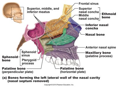
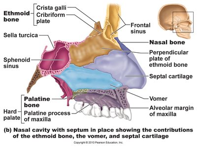
The bones forming the lateral wall and septum of the nasal cavity include the ethmoid, vomer, palatine, maxilla, and nasal bones. The conchae increase surface area for warming and humidifying air.
Superior and middle nasal conchae: Part of the ethmoid bone.
Inferior nasal concha: Separate bone.
Palatine bone: Forms part of the hard palate and lateral wall.
Vomer: Forms the inferior part of the nasal septum.
Paranasal Sinuses
Structure and Function of Paranasal Sinuses
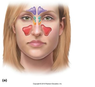
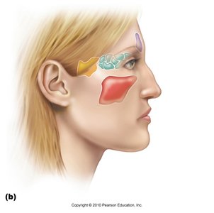
Paranasal sinuses are mucosa-lined, air-filled spaces in the frontal, maxillary, sphenoid, and ethmoid bones. They lighten the skull, enhance resonance of the voice, and help warm and humidify incoming air.
Frontal sinus
Maxillary sinus
Sphenoid sinus
Ethmoid sinus
Hyoid Bone
Structure and Function of the Hyoid Bone
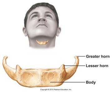
The hyoid bone is unique as it does not articulate with any other bone. It supports the tongue and provides attachment for muscles involved in swallowing and speech. It is horseshoe-shaped, consisting of a body and two pairs of horns (greater and lesser).
Greater horn: Larger, lateral projections.
Lesser horn: Smaller, medial projections.
Body: Central portion.
Summary Table: Cranial and Facial Bones
Bone | Type | Main Function |
|---|---|---|
Frontal | Cranial | Forehead, roof of orbits |
Parietal | Cranial | Superior/lateral skull |
Occipital | Cranial | Posterior skull, cranial base |
Temporal | Cranial | Inferior/lateral skull, cranial floor |
Sphenoid | Cranial | Base of middle cranial fossa |
Ethmoid | Cranial | Roof of nasal cavity, anterior cranial fossa |
Mandible | Facial | Lower jaw, holds teeth |
Maxilla | Facial | Upper jaw, hard palate |
Zygomatic | Facial | Cheekbone |
Nasal | Facial | Bridge of nose |
Lacrimal | Facial | Medial orbit, lacrimal sac |
Palatine | Facial | Hard palate, nasal cavity |
Vomer | Facial | Nasal septum |
Inferior nasal concha | Facial | Nasal cavity |
