 Indietro
The Integumentary System: Structure and Function
Guida di studio - Note intelligenti
 Appunti personalizzati basati sui tuoi materiali, ampliati con definizioni chiave, esempi e contesto.
Integumentary System Overview
Introduction to the Skin
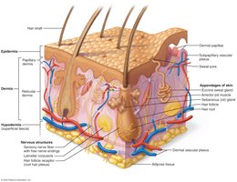
The skin is the largest organ of the human body, accounting for approximately 7% of total body weight. It varies in thickness and is composed of two main layers: the epidermis and the dermis. Beneath these lies the hypodermis, which, while not technically part of the integumentary system, shares several properties with the skin.
Epidermis: Outermost protective layer.
Dermis: Middle supportive layer containing connective tissue, blood vessels, and nerves.
Hypodermis: Deepest layer, primarily areolar and adipose tissue, anchors skin and provides insulation.
Functions of the Skin and Hypodermis
The skin and hypodermis perform several vital functions essential for homeostasis and protection:
Protection: Shields underlying tissues from mechanical damage, chemicals, water loss, and UV radiation.
Body Temperature Regulation: Capillary networks and sweat glands help regulate heat loss.
Excretion: Sweat glands excrete urea, salts, and water.
Vitamin D Production: Epidermal cells synthesize vitamin D when exposed to UV radiation.
Sensory Reception: Contains sensory receptors for touch, pain, and temperature.
Structure of the Epidermis
Cell Types in the Epidermis
The epidermis is a keratinized stratified squamous epithelium composed of four main cell types:
Keratinocytes: Most abundant; produce keratin, antibiotics, and enzymes; dead at the surface.
Melanocytes: Located in the basal layer; produce the pigment melanin.
Tactile Epithelial Cells: Basal layer; associated with sensory nerve endings.
Dendritic Cells: Stratum spinosum; immune function, macrophage-like.
Layers of the Epidermis
The epidermis is organized into distinct layers, each with specialized functions:
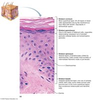
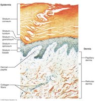
Stratum Basale (Germinativum): Deepest, actively dividing cells, contains melanocytes and tactile cells.
Stratum Spinosum: Several layers of keratinocytes, contains dendritic cells, resists tension.
Stratum Granulosum: Keratinocytes with keratohyaline and lamellar granules; cells begin to die above this layer.
Stratum Lucidum: Present only in thick skin (palms, soles); few rows of flat, dead keratinocytes.
Stratum Corneum: Outermost, thick layer of dead keratinocytes; protects against abrasion and penetration.
Dermis
Structure and Layers of the Dermis
The dermis is a strong, flexible connective tissue layer that supports and nourishes the epidermis. It contains two main layers:
Papillary Dermis: Superficial, contains dermal papillae that increase surface area for exchange and form epidermal ridges (fingerprints).
Reticular Dermis: Deeper, makes up 80% of dermal thickness, dense irregular connective tissue with collagen fibers for strength and elasticity.
Dermal Modifications
Specialized structures in the dermis include:
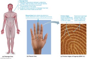
Flexure Lines: Deep creases in palms, wrists, soles, fingers, and toes.
Friction Ridges: Form fingerprints, enhance grip.
Vascular Plexuses: Networks of blood vessels important for temperature regulation.
Hypodermis
Structure and Function
The hypodermis (superficial fascia) lies deep to the skin and consists mainly of areolar and adipose connective tissue. It anchors the skin to underlying structures and provides insulation. The distribution of adipose tissue varies between males and females.
Skin Color
Pigments Contributing to Skin Color
Skin color is determined by three main pigments:
Melanin: Brown to black pigment produced by melanocytes; most significant determinant of skin color.
Carotene: Yellow-orange pigment from dietary sources like carrots and tomatoes.
Hemoglobin: Red pigment in blood; visible in individuals with little melanin.
Accessory Structures of the Skin
Nails
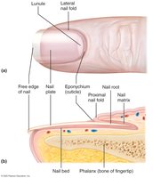
Nails are scalelike modifications of the epidermis composed of hard keratin. They protect the distal phalanx and enhance fine touch sensation.
Parts of the Nail: Free edge, nail plate, root, nail folds, and eponychium (cuticle).
Hair
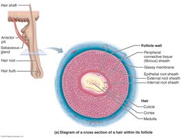
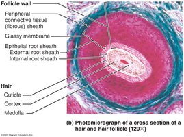
Hair consists of flexible strands of dead, keratinized cells. It is composed of three concentric layers:
Medulla: Central core.
Cortex: Surrounds the medulla.
Cuticle: Outermost layer.
Hair Follicles and Associated Structures
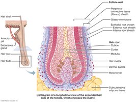
Hair follicles extend from the epidermis into the dermis. The hair bulb is the expanded base, surrounded by a root plexus of sensory nerves. The follicle wall consists of several layers, including the peripheral connective tissue sheath, glassy membrane, and epithelial root sheath. The arrector pili muscle is a bundle of smooth muscle that causes hair to stand erect when contracted.
Types and Growth of Hair
Vellus Hairs: Fine, body hairs of women and children.
Terminal Hairs: Coarser, longer hair of the scalp, axillary, and pubic regions (develop at puberty).
Glands of the Skin
Sebaceous Glands
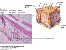
Sebaceous glands are simple alveolar glands found throughout the body except on the palms and soles. They secrete sebum, an oily substance that lubricates and waterproofs the skin and hair. Sebum is produced by holocrine secretion, where entire cells disintegrate to release their contents. Most sebaceous glands are associated with hair follicles.
Sweat Glands
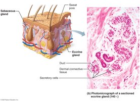
Sweat glands (sudoriferous glands) are widely distributed and play a key role in thermoregulation and excretion. Sweat is composed of 99% water, salts, and traces of metabolic wastes.
Eccrine Glands: Most numerous; produce true sweat for cooling the body.
Apocrine Glands: Confined to axillary, anal, and genital areas; produce a thicker, musky sweat that may function in mate attraction.
Modified Apocrine Glands: Include ceruminous (earwax) and mammary (milk) glands.
Summary Table: Layers of the Epidermis
Layer | Main Features | Location |
|---|---|---|
Stratum Corneum | Many layers of dead, keratinized cells; protection | Most superficial |
Stratum Lucidum | Clear, flat dead cells; only in thick skin | Palms, soles |
Stratum Granulosum | Keratohyaline and lamellar granules; cells begin to die | Middle layer |
Stratum Spinosum | Several layers; keratinocytes, dendritic cells | Above stratum basale |
Stratum Basale | Single row; mitotically active; melanocytes, tactile cells | Deepest |
