 Indietro
The Integumentary System: Structure, Function, and Clinical Aspects
Guida di studio - Note intelligenti
 Appunti personalizzati basati sui tuoi materiali, ampliati con definizioni chiave, esempi e contesto.
The Integumentary System
Overview and Components
The integumentary system is the body's largest organ system, serving as the primary barrier between the internal environment and the external world. It includes the skin and its derivatives, which perform vital protective and regulatory functions.
Skin: The main organ, consisting of two primary layers (epidermis and dermis) and an underlying subcutaneous layer (hypodermis).
Sudoriferous (sweat) glands: Involved in thermoregulation and excretion.
Sebaceous (oil) glands: Secrete sebum for lubrication and protection.
Hairs: Provide protection and sensory input.
Nails: Protect distal phalanges and aid in manipulation.
Subcutaneous tissue (hypodermis): Anchors skin, provides insulation and shock absorption.
Functions of the Skin
Protection: Acts as a barrier against mechanical injury, pathogens, and harmful substances.
Body Temperature Regulation: Maintains homeostasis through sweat production and blood flow regulation.
Cutaneous Sensations: Contains sensory receptors for touch, pain, and temperature.
Metabolic Functions: Synthesizes vitamin D, activates hormones, and aids in collagen turnover.
Blood Reservoir: Stores up to 5% of the body's blood volume.
Excretion: Eliminates nitrogenous wastes and excess salts via sweat.
Skin Structure
Layers of the Skin
The skin is composed of two main layers and an associated subcutaneous layer:
Epidermis: Outermost, avascular, composed of keratinized stratified squamous epithelium.
Dermis: Underlies the epidermis, vascular, composed mainly of dense connective tissue.
Subcutaneous tissue (Hypodermis): Not part of the skin proper; consists mostly of adipose tissue and some areolar connective tissue, providing insulation and anchoring the skin to underlying structures.
The Epidermis
Cell Types
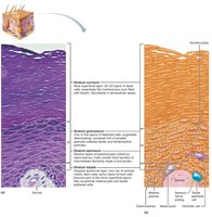
Keratinocytes: Produce keratin, the protein that gives skin its strength and waterproofing properties. Continuously renewed every 25–45 days.
Melanocytes: Produce melanin pigment, which protects against UV radiation.
Dendritic (Langerhans) cells: Immune cells that patrol the epidermis and activate immune responses.
Tactile epithelial (Merkel) cells: Sensory receptors for touch, located at the epidermal-dermal junction.
Layers (Strata) of the Epidermis
Stratum basale: Deepest, single row of mitotically active stem cells; contains melanocytes.
Stratum spinosum: Several layers thick; contains keratinocytes with pre-keratin filaments and abundant dendritic cells.
Stratum granulosum: 4–6 layers of flattened cells; keratinization begins, and lamellar granules provide water resistance.
Stratum lucidum: Thin, clear layer found only in thick skin (e.g., palms, soles); consists of dead keratinocytes.
Stratum corneum: 20–30 layers of dead, keratinized cells; provides the main barrier function.
The Dermis
Structure and Cell Types
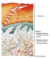
The dermis is a strong, flexible connective tissue layer housing blood vessels, nerves, glands, and hair follicles. It consists of two layers:
Papillary dermis: Superficial, areolar connective tissue with dermal papillae that project into the epidermis.
Reticular dermis: Deeper, dense irregular connective tissue containing collagen and elastic fibers.
Papillary Dermis
Contains dermal papillae, which increase surface area for exchange of gases, nutrients, and waste products.
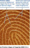
Forms friction ridges in thick skin, contributing to fingerprints and enhancing grip.
Reticular Dermis
Makes up the majority of dermal thickness.
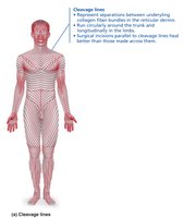
Contains cleavage (tension) lines, which are important for surgical incisions as they heal better when parallel to these lines.
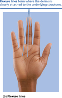
Flexure lines occur near joints where the dermis is tightly secured to underlying structures, forming visible creases.
Skin Color
Pigments Determining Skin Color
Melanin: Produced by melanocytes; protects DNA from UV damage. Varies in amount and type, resulting in different skin colors.
Carotene: Yellow to orange pigment, most visible in palms and soles; can be converted to vitamin A.
Hemoglobin: Oxygenated pigment in red blood cells; gives fair skin a pinkish hue due to transparency of the epidermis.
Alterations in skin color can indicate disease (e.g., cyanosis, jaundice, pallor, erythema, bruising).
Hair
Structure and Function
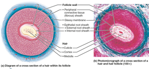
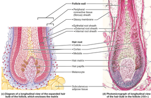
Hair is composed of dead, keratinized cells and serves protective and sensory functions. It is absent from palms, soles, lips, nipples, and parts of external genitalia.
Functions: Sensing insects, protecting scalp, reducing heat loss, shielding from sunlight, filtering air (nose), and protecting eyes (eyelashes).
Structure of a Hair
Root: Located within the scalp; keratinization ongoing.
Shaft: Projects above the skin; keratinization complete.
Three layers of the hair shaft:
Medulla: Central core with large cells and air spaces.
Cortex: Several layers of flattened cells surrounding the medulla.
Cuticle: Outermost layer of overlapping single cells.
Pigmentation: Determined by melanins; red hair contains pheomelanin; gray/white hair results from decreased melanin and air bubbles in the shaft.
Types and Growth of Hair
Vellus hair: Fine, pale body hair of children and adult females.
Terminal hair: Coarse, long hair found on scalp, eyebrows, and, after puberty, in axillary and pubic regions, and on the face and neck of males.
Hair growth is influenced by nutrition and hormones; follicles cycle between active and resting phases.
Nails
Structure and Function
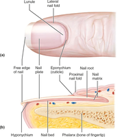
Nails are scale-like modifications of the epidermis containing hard keratin. They protect the distal phalanges and aid in manipulation and fine touch.
Skin Glands
Sweat Glands
Eccrine sweat glands: Most numerous; secrete watery sweat for thermoregulation.
Apocrine sweat glands: Found in axillary and anogenital areas; secrete a thicker, milky secretion.
Modified apocrine glands:
Ceruminous glands: Produce earwax.
Mammary glands: Secrete milk.
Sebaceous (Oil) Glands
Secrete sebum, an oily substance that lubricates skin and hair and has antibacterial properties.
Clinical Aspects: Skin Cancer and Burns
Skin Cancer
Skin cancer is primarily caused by UV exposure and chronic irritation. Early detection is crucial for successful treatment. The three major types are:
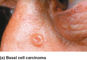
Basal cell carcinoma: Most common and least malignant; arises from stratum basale; slow-growing and rarely metastasizes.
Squamous cell carcinoma: Arises from keratinocytes of the stratum spinosum; can metastasize if not treated.
Melanoma: Cancer of melanocytes; highly metastatic and resistant to treatment; most dangerous form.
ABCDE Rule for Early Detection:
A: Asymmetry
B: Border irregularity
C: Color variation
D: Diameter > 6 mm
E: Evolution (change over time)
Burns
Burns are classified by depth and extent of tissue damage. The immediate threat is dehydration and electrolyte imbalance. Severity is assessed using the "rule of nines."
First-degree burns: Affect only the epidermis; cause redness and pain.
Second-degree burns: Involve the epidermis and upper dermis; cause blisters.
Third-degree burns: Destroy the entire thickness of the skin; area may appear white, red, or blackened; not painful due to nerve destruction; require skin grafting.
Burns are considered critical if:
>25% of body has second-degree burns
>10% of body has third-degree burns
Face, hands, or feet bear third-degree burns
Treatment includes debridement, antibiotics, temporary coverings, and skin grafts.
