 Indietro
The Integumentary System: Structure, Function, and Clinical Relevance
Guida di studio - Note intelligenti
 Appunti personalizzati basati sui tuoi materiali, ampliati con definizioni chiave, esempi e contesto.
The Integumentary System
Overview and Components
The integumentary system is the body's most visible organ system, comprising the skin and its accessory structures. It serves as a protective covering and plays vital roles in sensation, thermoregulation, and metabolic processes.
Skin (Integument): The largest and heaviest organ, covering 1.5–2.0 m2 and accounting for about 15% of adult body weight.
Accessory Organs: Hair, nails, cutaneous receptors (sensory structures), and cutaneous glands (sweat, sebaceous, ceruminous, mammary).
Clinical Relevance: Inspection of the skin, hair, and nails can reveal systemic diseases such as anemia, liver disorders, and heart failure.
Dermatology: The medical specialty focused on the integumentary system.
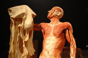
Functions of the Integumentary System
Barrier Functions: Protects against trauma, infection, water loss, UV radiation, and chemical exposure. The epidermis is packed with keratin and connected by strong desmosomes, forming a durable barrier.
Sensation: The skin is the most extensive sense organ, containing nerve endings that detect heat, cold, touch, pressure, vibration, and injury. Areas such as the face, palms, and soles are especially sensitive.
Vitamin D Synthesis: The epidermis initiates vitamin D synthesis, which is completed in the liver and kidneys.
Thermoregulation: Thermoreceptors and blood vessel dilation/constriction help regulate body temperature. Sweat glands provide evaporative cooling.
Excretion: Sweat glands excrete small amounts of lactic acid, electrolytes, urea, and some toxins.
Structure of the Skin
Skin Layers
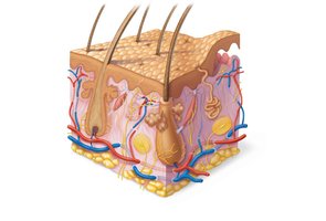
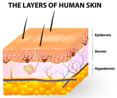
The skin consists of three main layers, each with distinct structures and functions:
Epidermis: Outermost layer, composed of keratinized stratified squamous epithelium. Lacks blood vessels and relies on diffusion from the underlying dermis.
Dermis: Middle layer, made of connective tissue (loose areolar and dense irregular). Contains blood vessels, nerves, hair follicles, and glands.
Hypodermis (Subcutaneous Tissue): Not technically part of the skin but closely associated. Composed of adipose and areolar tissue, providing insulation, energy storage, and shock absorption.
Thick vs. Thin Skin
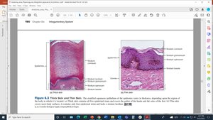
Skin is classified as thick or thin based on the number of epidermal layers and the thickness of the epidermis, not the entire integument.
Thick Skin: Found on palms, soles, and surfaces subject to abrasion. Contains a thick stratum corneum and a stratum lucidum. Lacks hair follicles and sebaceous glands. Thickness: 0.4–0.6 mm.
Thin Skin: Covers most of the body. Lacks a stratum lucidum and has a thinner stratum corneum. Contains hair follicles, sebaceous, and sweat glands. Thickness: 0.075–0.150 mm.
The Epidermis
Cell Types in the Epidermis
Stem Cells: Undifferentiated cells in the stratum basale that divide to produce keratinocytes.
Keratinocytes: Most abundant cells; produce keratin for strength and water resistance.
Melanocytes: Located in the stratum basale; synthesize melanin (eumelanin and pheomelanin) to protect against UV radiation.
Tactile (Merkel) Cells: Sensory receptors for light touch, associated with nerve endings.
Dendritic (Langerhans) Cells: Immune cells that guard against pathogens and alert the immune system.
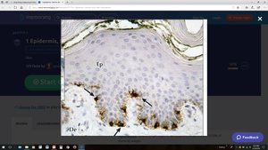
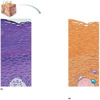
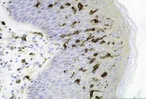
Layers (Strata) of the Epidermis
The epidermis is organized into four or five layers, depending on skin thickness:
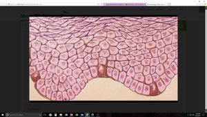
Stratum Basale: Deepest layer; single row of stem cells, melanocytes, and tactile cells. Site of cell division and initial vitamin D synthesis.
Stratum Spinosum: 8–10 layers of keratinocytes and dendritic cells; cells appear spiny due to desmosomes.
Stratum Granulosum: 3–5 layers of flattened keratinocytes with keratohyalin granules; cells begin keratinization and lose nuclei/organelles.
Stratum Lucidum: Only in thick skin; 2–3 layers of dead, translucent keratinocytes containing eleidin.
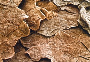
Stratum Corneum: Outermost layer; 5–30 layers of dead, keratinized cells providing a durable, water-resistant barrier.
The Dermis
Structure and Composition
The dermis is a connective tissue layer beneath the epidermis, providing structural support and housing most skin appendages.
Thickness: 0.2 mm (eyelids) to 4 mm (palms/soles).
Composition: Mainly collagen, with elastic and reticular fibers, fibroblasts, blood vessels, nerves, hair follicles, and glands.
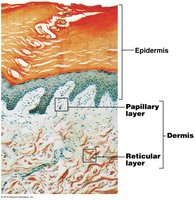
Dermal Papillae: Peg-like projections that increase surface area for nutrient exchange and anchor the epidermis.
Dermal Zones
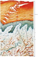
Papillary Layer: Superficial; composed of loose areolar connective tissue. Contains capillaries, lymphatics, nerve endings, and Meissner corpuscles (touch receptors).
Reticular Layer: Deep; dense irregular connective tissue with collagen and elastin fibers. Contains hair follicles, glands, blood vessels, and sensory receptors (Pacinian corpuscles, Ruffini endings).
Dermal Modifications
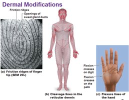
Friction Ridges: Form fingerprints and enhance grip.
Cleavage Lines: Correspond to collagen fiber orientation; important for surgical incisions to minimize scarring.
Flexure Lines: Dermal folds at joints, allowing skin to move with underlying structures.
Wrinkles, Stretch Marks, Scars: Result from aging, rapid stretching, or injury.
Moles, Freckles, Birthmarks: Localized concentrations of melanin or vascular changes.
The Hypodermis (Subcutaneous Tissue)
Structure and Function
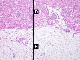
The hypodermis lies beneath the dermis and is composed of adipose and areolar connective tissue. It is not technically part of the skin but is functionally important.
Functions: Fat storage, insulation, shock absorption, and anchoring skin to underlying tissues.
Clinical Note: Site for subcutaneous injections.
Sex Differences: Thicker in females, contributing to body contours and energy reserves.
Skin Color and Pigmentation
Melanin and Its Role
Melanin is the primary determinant of skin color, produced by melanocytes in the stratum basale and transferred to keratinocytes. It protects DNA from UV radiation.
Types of Melanin: Eumelanin (brown-black) and pheomelanin (reddish-yellow).
Variation: All humans have similar numbers of melanocytes; differences in skin color arise from the amount, type, and distribution of melanin.
Melanosomes: Melanin-containing organelles that cluster above keratinocyte nuclei to shield DNA from UV damage.
Summary Table: Layers of the Skin
Layer | Main Tissue Type | Key Features |
|---|---|---|
Epidermis | Keratinized stratified squamous epithelium | Protection, water resistance, sensory input |
Dermis | Connective tissue (areolar & dense irregular) | Support, vascular supply, houses appendages |
Hypodermis | Adipose & areolar connective tissue | Insulation, energy storage, shock absorption |
