 Indietro
The Integumentary System: Structure, Function, and Clinical Relevance
Guida di studio - Note intelligenti
 Appunti personalizzati basati sui tuoi materiali, ampliati con definizioni chiave, esempi e contesto.
The Integumentary System
Overview
The integumentary system is the body's largest organ system, providing protection, sensation, and regulation. It consists of the skin, sweat glands, oil glands, hair, nails, and subcutaneous tissue. This system plays a vital role in maintaining homeostasis and defending against environmental hazards.
Structure of the Skin
Layers of the Skin
Epidermis: Outermost protective shield, composed of keratinized stratified squamous epithelium; avascular.
Dermis: Underlies the epidermis, primarily dense connective tissue; vascular and provides structural strength.
Subcutaneous Tissue (Hypodermis): Not technically part of the skin, but functionally related. Composed mostly of adipose tissue and some areolar connective tissue. Functions include anchoring skin to underlying structures, shock absorption, and insulation.
The Epidermis
Cell Types
Keratinocytes: Produce keratin, a protein that provides protective properties. Major cell type; replaced every 25-45 days.
Melanocytes: Produce melanin pigment, which protects against UV damage. Located in the basal layer.
Dendritic (Langerhans) Cells: Macrophages that patrol the epidermis and activate the immune system.
Tactile (Merkel) Cells: Sensory receptors for touch, located at the epidermal-dermal junction.
Layers (Strata) of the Epidermis
Stratum Basale: Deepest layer; single row of mitotically active stem cells. Also called stratum germinativum.
Stratum Spinosum: Several layers thick; contains pre-keratin filaments and abundant dendritic cells.
Stratum Granulosum: Four to six cell layers; keratinization begins, and cells accumulate keratohyaline and lamellar granules.
Stratum Lucidum: Present only in thick skin (palms, soles); thin, clear layer of dead keratinocytes.
Stratum Corneum: Outermost layer; 20-30 rows of dead, keratinized cells providing protection and water resistance.
Note: Thick skin contains all five layers; thin skin lacks the stratum lucidum.
The Dermis
Structure and Function
The dermis is a strong, flexible connective tissue layer containing fibroblasts, macrophages, mast cells, and white blood cells. It houses nerves, blood vessels, lymphatics, hair follicles, and glands.
Layers of the Dermis
Papillary Dermis: Superficial layer of areolar connective tissue with dermal papillae containing capillaries, nerve endings, and touch receptors. Forms friction ridges (fingerprints) in thick skin.
Reticular Dermis: Deep layer (~80% of dermal thickness); dense irregular connective tissue with collagen and elastic fibers. Provides strength, resiliency, and hydration. Contains cleavage (tension) lines and flexure lines.
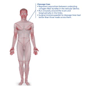
Cleavage lines represent separations between underlying collagen fiber bundles in the reticular dermis. Surgical incisions parallel to these lines heal better than those made across them.
Clinical Relevance
Striae (Stretch Marks): Dermal tears from extreme stretching.
Blisters: Fluid-filled pockets separating epidermal and dermal layers due to trauma.
Skin Color
Pigments Determining Skin Color
Melanin: Produced by melanocytes; protects DNA from UV damage. Varies in amount and form, causing different skin tones.
Carotene: Yellow-orange pigment, most visible in palms and soles; can be converted to vitamin A.
Hemoglobin: Gives pinkish hue to fair skin due to low melanin levels.
Clinical Correlations
Cyanosis: Blue skin from low oxygenation.
Pallor: Pale skin from anemia or stress.
Erythema: Redness from fever, inflammation, or allergy.
Jaundice: Yellow skin from liver disorders.
Bruises: Blood leakage under skin; color changes with healing.
Hair
Structure and Function
Functions: Sensory detection, protection from trauma, heat loss, and sunlight; eyelashes and nose hairs filter debris.
Structure: Dead, keratinized cells produced by hair follicles. Regions include root (in scalp) and shaft (above scalp). Shaft has medulla, cortex, and cuticle layers.
Pigmentation: Melanocytes in follicles produce pigments; red hair has pheomelanin; gray/white hair results from decreased melanin and air bubbles.
Hair Follicle Anatomy
Hair Bulb: Deep end of follicle; contains hair matrix (actively dividing cells) and papilla (nutrient supply).
Arrector Pili: Smooth muscle causing "goosebumps."
Types and Growth
Vellus Hair: Fine, pale body hair.
Terminal Hair: Coarse, long hair (scalp, eyebrows, axillary, pubic regions).
Growth Cycles: Alternating active and resting phases; scalp hair grows for years, eyebrows for months.
Clinical Relevance
Hirsutism: Excessive hair growth in females, often due to androgen excess.
Baldness: Age-related or genetic (male pattern baldness); follicles respond to androgens with shorter growth cycles.
Nails
Structure and Function
Composition: Scale-like modifications of the epidermis with hard keratin.
Parts: Root (embedded), nail plate/body (visible), free edge, nail bed (under plate), nail matrix (growth area), nail folds, cuticle (eponychium), hyponychium (under free edge).
Clinical Indicators: Nail color and shape can indicate systemic diseases (e.g., yellow nails for respiratory disorders, spoon nails for iron deficiency).
Sweat and Oil Glands
Sweat Glands
Eccrine (Merocrine) Glands: Most numerous; secrete watery sweat for thermoregulation; found on palms, soles, forehead.
Apocrine Glands: Found in axillary and anogenital areas; secrete viscous sweat into hair follicles; function begins at puberty.
Modified Apocrine Glands: Ceruminous (earwax) and mammary (milk) glands.
Sebaceous (Oil) Glands
Distribution: All over body except palms and soles; usually associated with hair follicles.
Secretion: Sebum (oily, bactericidal, softens skin and hair); activity increases at puberty.
Clinical: Acne (inflammation of sebaceous glands), cradle cap (seborrhea in infants).
Functions of the Skin
Main Functions
Protection: Chemical (antimicrobial, acid mantle, melanin), physical (keratinized cells, glycolipids), and biological (dendritic cells, macrophages) barriers.
Body Temperature Regulation: Insensible and sensible perspiration; vasodilation and vasoconstriction of dermal vessels.
Cutaneous Sensation: Sensory receptors for touch, pressure, pain, and temperature.
Metabolic Functions: Synthesis of vitamin D, activation of hormones, collagenase production.
Blood Reservoir: Holds up to 5% of blood volume; can be shunted as needed.
Excretion: Eliminates nitrogenous wastes, salt, and water through sweat.
Skin Disorders: Cancer and Burns
Skin Cancer
Basal Cell Carcinoma: Most common, least malignant; arises from stratum basale; high cure rate with excision.
Squamous Cell Carcinoma: Arises from stratum spinosum; can metastasize; good prognosis if treated early.
Melanoma: Cancer of melanocytes; highly metastatic and dangerous; treated with surgery and immunotherapy.
ABCD(E) Rule for Melanoma Detection: Asymmetry, Border irregularity, Color variation, Diameter >6mm, Evolution.
Burns
First Degree: Epidermal damage only; redness, swelling, pain.
Second Degree: Epidermal and upper dermal damage; blisters.
Third Degree: Full-thickness; gray-white, cherry red, or blackened skin; not painful (nerve endings destroyed); requires grafting.
Rule of Nines: Used to estimate fluid loss by dividing the body into sections, each representing 9% of total surface area.
Developmental Aspects
Embryonic: Epidermis from ectoderm, dermis and hypodermis from mesoderm; skin well developed by 4th month.
Fetal: Lanugo coat (fine hair) and vernix caseosa (waxy covering) protect fetus.
Infancy to Adulthood: Skin thickens, accumulates more subcutaneous fat, and sweat and sebaceous gland activity increases at puberty.
Summary Table: Layers of the Epidermis
Layer | Main Features | Location |
|---|---|---|
Stratum Corneum | 20-30 layers of dead, keratinized cells | Outermost |
Stratum Lucidum | Clear, dead cells (only in thick skin) | Palms, soles |
Stratum Granulosum | Keratinization begins, granules present | Middle |
Stratum Spinosum | Spiny cells, desmosomes, dendritic cells | Above basal |
Stratum Basale | Mitotically active stem cells, melanocytes | Deepest |
