 Indietro
The Integumentary System: Structure, Function, and Associated Tissues
Guida di studio - Note intelligenti
 Appunti personalizzati basati sui tuoi materiali, ampliati con definizioni chiave, esempi e contesto.
The Integumentary System
Definition and Components
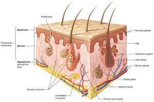
The integumentary system consists of the skin, hair, glands, and nails. The skin is the largest organ in the body, accounting for 10-15% of total body weight. It serves as the primary interface between the body and the external environment.
Functions of the Integumentary System
Protection: The skin provides a durable, flexible barrier against mechanical trauma, pathogens, and environmental factors such as UV radiation and water loss.
Sensation: Numerous sensory receptors in the skin detect changes in the internal and external environment, aiding homeostasis.
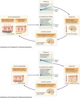
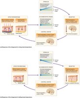
Thermoregulation: The skin regulates body temperature through negative feedback loops involving sweat glands and blood vessels.
Excretion: Sweat eliminates small amounts of waste products.
Vitamin D Synthesis: The skin initiates vitamin D production upon exposure to UV radiation, which is essential for calcium absorption and bone health.
Thermoregulation Mechanisms
Thermoregulation involves physiological responses to rising or falling body temperature, primarily through sweat production and blood vessel dilation or constriction.
Structure of the Skin
Keratinocytes and Epidermal Layers
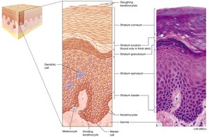
Keratinocytes make up 95% of epidermal cells, producing keratin for mechanical strength. These cells are organized into distinct layers, or strata, each with specialized functions and cell types.
Layers of the Epidermis (Deep to Superficial)
Stratum Basale: Single layer of stem cells, most metabolically active, produces vitamin D precursor.
Stratum Spinosum: Thickest layer, metabolically active, helps produce vitamin D.
Stratum Granulosum: Releases lipid-based substances, creating hydrophobic skin properties and isolating cells.
Stratum Lucidum: Narrow, clear layer of dead keratinocytes, found only in thick skin.
Stratum Corneum: Several layers of dead, flattened keratinocytes, which eventually slough off.
Epidermal Growth and Cell Sloughing
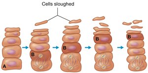
Keratinocytes undergo mitosis in the stratum basale and migrate upward, eventually dying and sloughing off in the stratum corneum.
Other Cells of the Epidermis
Dendritic (Langerhans) Cells: Immune phagocytes in the stratum spinosum.
Merkel Cells: Sensory receptors for light touch, found in the stratum basale.
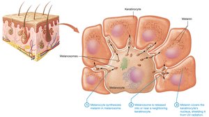
Melanocytes: Produce melanin pigment, located in the stratum basale.
Thick vs. Thin Skin
Thick Skin: Found on palms and soles, contains all five strata, lacks hair follicles, has many sweat glands.
Thin Skin: Found elsewhere, lacks stratum lucidum, has thinner strata, contains hair follicles, sebaceous glands, and sweat glands.
The Dermis
Layers of the Dermis
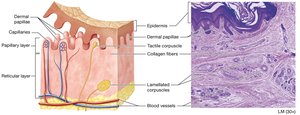
Papillary Layer: Loose connective tissue, contains dermal papillae with capillaries and Meissner corpuscles.
Reticular Layer: Dense irregular connective tissue, contains collagen and elastic fibers, blood vessels, glands, hair follicles, and Pacinian corpuscles.
Fingerprints and Epidermal Ridges
Formation and Function
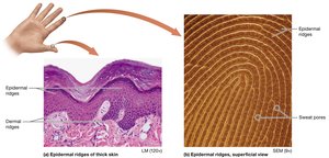
Epidermal ridges are visible lines on thick skin, formed by prominent dermal papillae and collagen fibers. These ridges enhance grip and create unique fingerprint patterns (loops, arches, whorls) via sweat pores.
Skin Pigmentation
Melanin
Melanin is produced by melanocytes in the stratum basale and ranges from orange-red to black. It protects keratinocyte DNA from UV-induced mutations and regulates vitamin D synthesis. Melanin concentration varies across the body, and its synthesis increases with UV exposure.
Carotene and Hemoglobin
Carotene: Yellow-orange pigment from diet, accumulates in the stratum corneum, especially visible in thick skin.
Hemoglobin: Iron-containing protein in red blood cells, gives skin a pink tone in lighter-skinned individuals due to vascular dermis and translucent epidermis.
Accessory Structures of the Integument
Hair
Functions: Thermoregulation (minor in humans), protection from foreign objects and UV radiation, sensation.
Structure: Hair shaft (dead, keratinized cells), root (living cells), matrix (mitotic keratinocytes), follicle (epithelial and dermal root sheaths), arrector pili muscle (piloerection).
Regions: Medulla (innermost, thick hairs), cortex (superficial to medulla), cuticle (outermost).
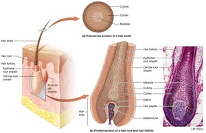
Hair Pigmentation
Lanugo: Thin, nonpigmented fetal hair.
Terminal Hair: Thick, coarse, pigmented hair (replaces vellus hair after puberty).
Vellus Hair: Thin, nonpigmented hair.
Melanin determines hair color; red hair contains a pigment with iron.
Nails
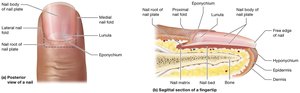
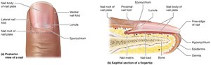
Composed of stratified squamous epithelium with hard keratin.
Eponychium (cuticle): Stratum corneum only.
Hyponychium: Attaches distal nail to nail bed, also stratum corneum.
Nails lack melanocytes.
Functions: Protect underlying tissue, prevent trauma, aid in gripping.
Glands
Sweat (Sudoriferous) Glands: Eccrine (most prevalent, thermoregulation), Apocrine (armpits, anal area, areolae, protein-rich, active after puberty), Ceruminous (ear wax), Mammary (milk production).
Sebaceous Glands: Produce sebum (oil), holocrine secretion, hydrophobic barrier, found throughout body except palms and soles.
Connective Tissue Example: Reticular Tissue
Structure and Function
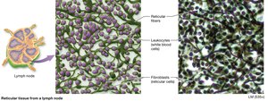
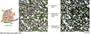
Reticular tissue is a type of connective tissue found in lymph nodes. It consists of reticular fibers, leukocytes (white blood cells), and fibroblasts (reticular cells). This tissue forms a supportive framework for immune cells.
Summary Table: Types of Skin Glands
Gland Type | Location | Product | Function |
|---|---|---|---|
Eccrine Sweat | Most skin areas | Sweat (99% water) | Thermoregulation |
Apocrine Sweat | Armpits, anal area, areolae | Protein-rich sweat | Active after puberty |
Ceruminous | Ear canal | Cerumen (ear wax) | Lubrication, protection |
Mammary | Breasts | Milk | Nourishment of infants |
Sebaceous | Most skin (not palms/soles) | Sebum (oil) | Hydrophobic barrier |
Key Terms and Concepts
Keratin: Fibrous protein for mechanical strength
Melanin: Pigment for UV protection
Desmosome: Cell junction for mechanical stability
Tight Junction: Cell junction preventing passage of substances
Stratum: Layer of the epidermis
Holocrine Secretion: Release of product via cell rupture
Sample Equations
Vitamin D Synthesis (simplified):
Calcium Absorption (regulated by Vitamin D):
Additional info:
Reticular tissue is part of the lymphatic system and supports immune cell function.
Fingerprints are unique to each individual and are used for identification.
Holocrine secretion is unique to sebaceous glands among skin glands.
