 Indietro
The Integumentary System: Structure, Function, and Clinical Relevance
Guida di studio - Note intelligenti
 Appunti personalizzati basati sui tuoi materiali, ampliati con definizioni chiave, esempi e contesto.
The Integumentary System
Overview and Components
The integumentary system is the body's largest organ system, serving as the primary interface between the internal environment and the outside world. It consists of the skin, hair, nails, sweat glands, and sebaceous (oil) glands. These structures collectively provide protection, regulate temperature, and facilitate sensory perception.
Skin: The main organ, composed of multiple layers.
Hair: Provides protection and sensory input.
Nails: Protects the tips of fingers and toes.
Sweat glands: Regulate temperature and excrete wastes.
Sebaceous glands: Secrete oil to lubricate skin and hair.
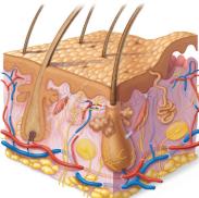
Skin Structure
Layers of the Skin
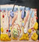
The skin is composed of two primary layers: the epidermis and the dermis. Beneath these lies the hypodermis (subcutaneous layer), which is not technically part of the skin but shares some functions.
Epidermis: Superficial, avascular layer made of epithelial tissue.
Dermis: Deeper, vascular layer made of connective tissue.
Hypodermis: Mostly adipose tissue; absorbs shock, insulates, and anchors skin to underlying structures.
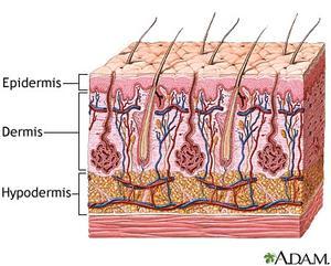
Epidermis
Cell Types in the Epidermis
The epidermis is primarily composed of keratinized stratified squamous epithelium and contains four main cell types:
Keratinocytes: Produce keratin, a protein that provides strength and waterproofing. Tightly connected by desmosomes.
Melanocytes: Produce melanin pigment, which protects against UV radiation.
Dendritic (Langerhans) cells: Immune cells that patrol the epidermis.
Tactile (Merkel) cells: Sensory receptors for touch.
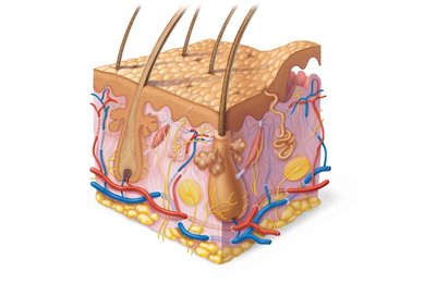
Layers of the Epidermis
The epidermis is organized into distinct layers (strata). Thick skin (palms, soles) has five layers; thin skin has four.
Stratum basale: Deepest layer; site of active mitosis and contains melanocytes.
Stratum spinosum: Several layers thick; contains keratinocytes, melanosomes, and dendritic cells.
Stratum granulosum: Cells flatten, organelles disintegrate, keratinization begins, lamellar granules accumulate.
Stratum lucidum: Only in thick skin; thin, translucent band of dead keratinocytes.
Stratum corneum: Outermost layer; 20–30 rows of dead, keratinized cells providing protection.
Dermis
Structure and Layers
The dermis is a strong, flexible connective tissue layer containing fibroblasts, macrophages, mast cells, and white blood cells. It houses nerves, blood vessels, lymphatic vessels, hair follicles, oil glands, and sweat glands.
Papillary layer: Superficial areolar connective tissue; contains dermal papillae with capillary loops, nerve endings, and touch receptors.
Reticular layer: Deep, dense fibrous connective tissue; provides strength, stretch, and water binding. Contains cleavage (tension) lines and flexure lines.

Skin Color
Pigments Contributing to Skin Color
Skin color is determined by three main pigments:
Melanin: Produced by melanocytes; shields DNA from UV radiation. Two forms: reddish yellow and brownish black.
Carotene: Yellow to orange pigment; accumulates in stratum corneum and hypodermis.
Hemoglobin: Gives pinkish hue to fair skin due to lower melanin levels.
Clinical Significance of Skin Color
Alterations in skin color can indicate disease:
Cyanosis: Blue skin; low oxygenation.
Erythema: Redness; fever, hypertension, inflammation, allergy.
Pallor: Pale color; anemia, low blood pressure, fear, anger.
Jaundice: Yellow cast; liver disorders.
Bronzing: Inadequate steroid hormones (Addison’s disease).
Bruises: Clotted blood beneath skin.
Hair
Structure and Function
Hair consists of dead, keratinized cells and is produced by hair follicles. It serves to protect, sense, and regulate temperature.
Shaft: Visible part; keratinization complete.
Root: Within scalp; keratinization ongoing.
Medulla: Central core.
Cortex: Surrounds medulla.
Cuticle: Outer layer.

Hair Follicle
Hair bulb: Expanded area at deep end.
Hair follicle receptor: Sensory nerve endings.
Wall: Peripheral connective tissue sheath, glassy membrane, epithelial root sheath.
Hair matrix: Actively dividing area.
Arrector pili: Smooth muscle causing goose bumps.
Hair papilla: Capillaries supplying nutrients.
Types and Growth
Vellus hair: Fine, pale body hair.
Terminal hair: Coarse, long hair.
Alopecia: Hair thinning after age 40.
Male pattern baldness: Genetically determined, influenced by DHT.
Nails
Structure and Function
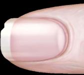
Nails are scale-like modifications of the epidermis containing hard keratin. They protect the tips of fingers and toes and consist of the free edge, nail plate, and root.
Nail bed: Epidermis under nail plate.
Nail matrix: Responsible for nail growth.
Nail folds: Skin folds overlapping nail border.
Eponychium: Cuticle.
Hyponychium: Area under free edge.
Lunule: White, thickened nail matrix.
Glands of the Skin
Sweat Glands
Eccrine (merocrine) sweat glands: Most numerous; abundant on palms, soles, forehead; function in thermoregulation; secrete sweat (99% water, salts, vitamin C, antibodies, wastes).
Apocrine sweat glands: Confined to axillary and anogenital areas; secrete viscous sweat; begin functioning at puberty; may act as sexual scent glands.
Modified apocrine glands: Ceruminous (earwax) and mammary (milk) glands.
Sebaceous (Oil) Glands
Widely distributed: Except palms and soles.
Develop from hair follicles: Secrete into hair follicles.
Inactive until puberty: Stimulated by hormones.
Secrete sebum: Oily, bactericidal, softens hair and skin.
Functions of the Skin
Protection
The skin acts as a barrier against microorganisms, abrasions, temperature extremes, and chemicals.
Chemical barrier: Sweat, sebum, defensins, acid mantle, melanin.
Physical barrier: Keratinized cells and glycolipids block water and substances.
Biological barrier: Dendritic cells and macrophages activate immune response.
Body Temperature Regulation
Insensible perspiration: 500 ml/day of unnoticeable sweat.
Sensible perspiration: Up to 12 L/day when overheated.
Cold environment: Dermal blood vessels constrict to conserve heat.
Cutaneous Sensation
Exteroreceptors: Respond to external stimuli (temperature, touch).
Free nerve endings: Sense pain.
Metabolic Functions
Vitamin D synthesis: Needed for calcium absorption.
Keratinocytes: Disarm carcinogens, activate hormones, produce collagenase.
Blood Reservoir
Holds up to 5% of blood volume: Can be shunted to other organs.
Excretion
Excretes nitrogenous wastes: Ammonia, urea, uric acid.
Sweating: Causes salt and water loss.
Clinical Conditions
Skin Cancer
Basal cell carcinoma: Most common, least malignant; affects stratum basale cells.
Squamous cell carcinoma: Second most common; involves keratinocytes of stratum spinosum.
Melanoma: Cancer of melanocytes; most dangerous, highly metastatic.
Burns
First-degree: Epidermal damage only; redness, edema, pain.
Second-degree: Epidermal and upper dermal damage; blisters.
Third-degree: Entire thickness of skin; gray-white, cherry red, or blackened; no pain (nerve endings destroyed); skin grafting required.
Rule of Nines
Used to estimate fluid loss and severity of burns. Body is divided into 11 sections, each representing 9% of surface area (except genitals, 1%).
Body Region | % of Surface Area |
|---|---|
Head and Neck | 9% |
Upper Limbs | 18% |
Trunk | 36% |
Lower Limbs | 36% |
Perineum | 1% |
Summary Table: Layers of the Skin
Layer | Main Features |
|---|---|
Stratum basale | Mitotic stem cells, melanocytes |
Stratum spinosum | Keratinocytes, melanosomes, dendritic cells |
Stratum granulosum | Keratinization, lamellar granules |
Stratum lucidum | Clear, dead keratinocytes (thick skin only) |
Stratum corneum | Dead, keratinized cells; protection |
Dermis | Papillary and reticular layers; nerves, vessels, glands |
Hypodermis | Adipose tissue; shock absorption, insulation |
