Indietro
The Integumentary System: Structure, Function, and Clinical Significance
Guida di studio - Note intelligenti
 Appunti personalizzati basati sui tuoi materiali, ampliati con definizioni chiave, esempi e contesto.
The Integumentary System
Overview and Functions
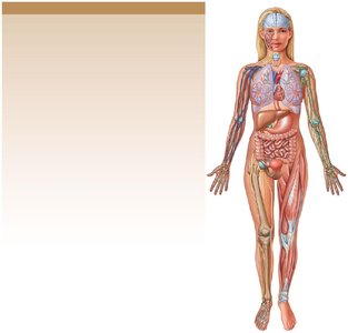
The integumentary system is the largest organ system in the human body, comprising approximately 16% of body weight and covering 1.5 to 2 m2 of surface area. It consists of the cutaneous membrane (skin) and accessory structures such as hair, glands, and nails. The system plays a vital role in protection, sensation, thermoregulation, and metabolic functions.
Protection: Shields underlying tissues from mechanical, chemical, and microbial threats.
Excretion: Removes salts, water, and organic wastes.
Thermoregulation: Maintains body temperature via blood flow and sweat production.
Vitamin D3 Synthesis: Essential for calcium metabolism.
Sensory Detection: Detects touch, pressure, pain, and temperature.
Immune Coordination: Responds to pathogens and cancers.
Structure of the Skin
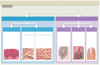
Cutaneous Membrane
The skin is composed of two primary layers: the epidermis (outer layer) and the dermis (inner layer). Beneath the dermis lies the subcutaneous layer (hypodermis), which anchors the skin to underlying tissues.
Epidermis: Stratified squamous epithelium; avascular.
Dermis: Connective tissue; contains blood vessels, nerves, and accessory structures.
Subcutaneous Layer: Loose connective tissue; stores fat and provides insulation.
Epidermis: Cell Types and Layers
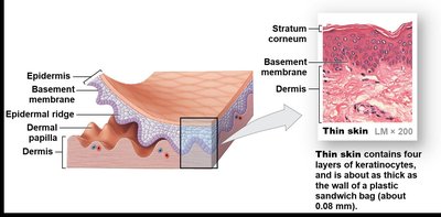
The epidermis is primarily composed of keratinocytes, which produce the protein keratin. It is organized into distinct layers (strata), with differences between thin skin and thick skin.
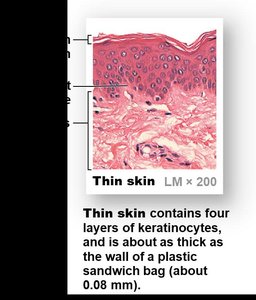
Thin Skin: Four layers of keratinocytes; covers most of the body.
Thick Skin: Five layers; found on palms and soles.
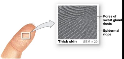
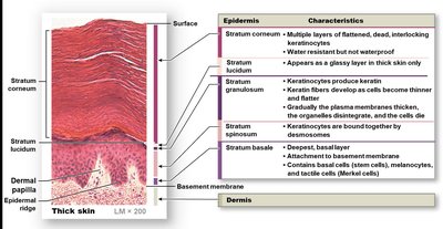
Stratum | Characteristics |
|---|---|
Stratum corneum | Multiple layers of flattened, dead, interlocking keratinocytes; water resistant |
Stratum lucidum | Appears as a glassy layer in thick skin only |
Stratum granulosum | Keratinocytes produce keratin; keratin fibers develop |
Stratum spinosum | Keratinocytes bound together by desmosomes |
Stratum basale | Deepest, basal layer; attachment to basement membrane; contains basal cells, melanocytes, and tactile cells |
Dermis: Layers and Functions
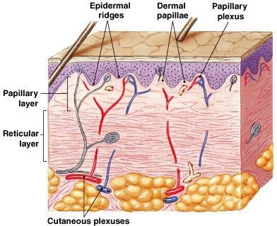
The dermis lies beneath the epidermis and provides structural support, nourishment, and sensory functions. It is divided into two layers:
Papillary Layer: Areolar tissue; contains capillaries, lymphatics, and sensory neurons; forms dermal papillae.
Reticular Layer: Dense irregular connective tissue; contains collagen and elastic fibers; houses accessory structures.
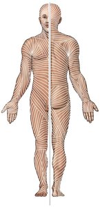
Tension Lines (Cleavage Lines)
Tension lines are formed by parallel bundles of collagen and elastic fibers in the dermis. They are clinically significant for surgical incisions, as cuts parallel to these lines heal better and with less scarring.
Skin Color
Pigments and Blood Flow
Skin color is determined by the presence of two main pigments and the oxygenation of blood:
Melanin: Produced by melanocytes; protects against UV radiation.
Carotene: Derived from diet; can be converted to vitamin A.
Hemoglobin: Oxygenated blood gives a reddish hue; deoxygenated blood can cause cyanosis (bluish skin).
Accessory Structures
Hair and Hair Follicles
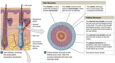
Hair is produced by follicles located in the dermis. It consists of keratinized cells and serves protective and sensory functions.
Structure: Medulla (core), cortex (thick layers), cuticle (outer layer).
Follicle: Internal and external root sheaths, glassy membrane, connective tissue sheath.
Glands of the Skin
The skin contains several types of exocrine glands:
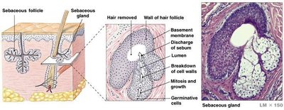
Sebaceous (Oil) Glands: Secrete sebum into hair follicles; lubricates and protects.
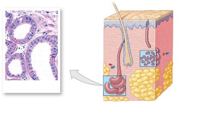
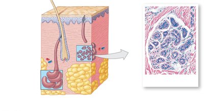
Apocrine Sweat Glands: Found in specific regions; produce odorous secretions.
Eccrine (Merocrine) Sweat Glands: Widely distributed; produce watery sweat for thermoregulation.
Other Glands: Mammary (milk) and ceruminous (earwax) glands.
Nails
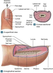
Nails protect the tips of fingers and toes and are composed of dead, keratinized cells. Their structure and growth are important indicators of health.
Nail Body: Visible portion covering the nail bed.
Nail Root: Site of nail production; lies close to bone.
Eponychium: Cuticle; protects the nail root.
Lunula: Pale crescent near the root.
Repair and Aging of the Skin
Repair Mechanisms
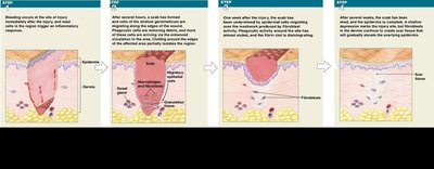
When the skin is injured, a series of steps occur to restore integrity:
Bleeding and inflammation
Formation of a scab
Macrophage activity to clean debris
Fibroblast proliferation and granulation tissue formation
Remodeling and scar formation
Aging Effects
Aging leads to structural and functional changes in the skin:
Thinning of the epidermis and dermis
Reduced immune response
Decreased vitamin D3 production
Decline in melanocyte and glandular activity
Slower repair rates
Integration with Other Body Systems
The integumentary system interacts with other systems to maintain homeostasis, protect against environmental hazards, and support metabolic functions.