 Indietro
The Lymphatic and Immune Systems: Structure and Function
Guida di studio - Note intelligenti
 Appunti personalizzati basati sui tuoi materiali, ampliati con definizioni chiave, esempi e contesto.
The Lymphatic and Immune Systems
Overview and Functions
The lymphatic system is a network of vessels, nodes, and organs that plays a crucial role in maintaining fluid balance, absorbing dietary fats, and supporting immune defense. The immune system consists of structures and cells that protect the body against disease, many of which are part of the lymphatic system.
Transports interstitial fluid (lymph) from tissues back to the bloodstream.
Transports absorbed fats from the small intestine to the blood.
Transports immune cells to and from lymph nodes and the blood.
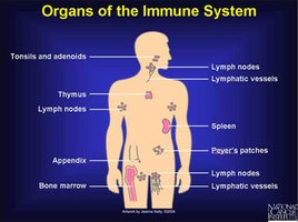
Major Structures of the Lymphatic System
Primary and Secondary Lymphatic Organs
The lymphatic system is composed of several key structures:
Lymphatic vessels: Transport lymph throughout the body.
Lymph nodes: Filter lymph and house lymphocytes.
Spleen: Filters blood, removes old red blood cells, and supports immune function.
Thymus: Site of T lymphocyte maturation.
Tonsils: Protect against pathogens entering through the mouth and throat.
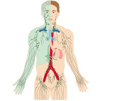
Lymphatic Circulation and Fluid Recovery
Relationship to the Cardiovascular System
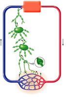
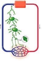
The lymphatic system works closely with the cardiovascular system to maintain fluid balance. Blood capillaries leak fluid into tissues, forming interstitial fluid. This fluid is collected by lymphatic capillaries and becomes lymph once inside the lymphatic vessels.
One-way system: Lymphatic vessels return fluid to the blood, preventing tissue swelling (edema).
Entry point: Lymphatic capillaries are closed-end tubes made of simple squamous epithelium.
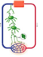
Lymphatic Capillaries and Valves
Structure and Function
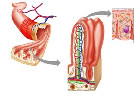
Lymphatic capillaries are highly permeable vessels that collect excess tissue fluid, fats, and microorganisms. Their walls are formed by overlapping endothelial cells that act as one-way valves, opening when interstitial fluid pressure increases.
Flaplike minivalves allow fluid entry but prevent backflow.
Lacteals: Specialized lymphatic capillaries in the small intestine that absorb dietary fats.
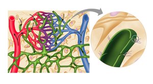
Lymphatic Collecting Vessels
Layers and Valves
Lymphatic collecting vessels drain lymph from capillaries and transport it toward lymph nodes and trunks. They have the same three layers as blood vessels but are much thinner:
Tunica intima: Endothelium, forms valves to prevent backflow.
Tunica media: Smooth muscle, helps propel lymph.
Tunica externa: Connective tissue.
Valves are essential because lymph is under low pressure and must be kept moving in one direction.
Lymph Nodes
Structure and Function
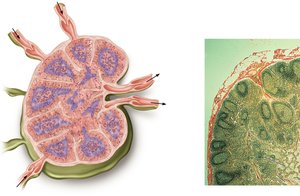
Lymph nodes are small, bean-shaped organs that filter lymph and house immune cells. There are about 500 lymph nodes in the human body, occurring alone or in clusters. Lymph passes through multiple nodes before returning to the bloodstream.
Afferent vessels: Bring lymph into the node.
Efferent vessels: Drain lymph away from the node.
Cortex: Outer region containing lymphocytes.
Medulla: Inner region with medullary sinuses and immune cells.
Why Lymph Nodes Swell
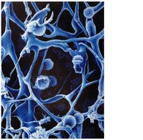
Accumulation of viruses and pathogens trapped in the node.
Increased number of lymphocytes fighting infection.
Excess fluid to dilute pathogens.
All of the above can contribute to lymph node swelling during illness.
Lymphatic Trunks and Ducts
Major Lymphatic Trunks
Lymphatic trunks are large vessels that drain lymph from major regions of the body. Examples include the jugular, subclavian, bronchomediastinal, intestinal, and lumbar trunks.
Lymphatic Ducts
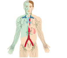
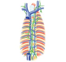
There are only two main lymphatic ducts:
Right lymphatic duct: Drains the right side of the head, neck, arm, and thoracic cavity into the right subclavian vein.
Thoracic duct: Drains the rest of the body into the left brachiocephalic vein.
Clinical Correlations
Lymphedema
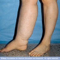
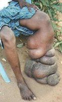
Lymphedema is swelling in a region of the body due to obstruction within the lymphatic structures. In the U.S., it is most commonly caused by removal or enlargement of lymph nodes. In other parts of the world, parasitic infection is a frequent cause.
Elephantiasis
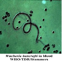
Elephantiasis is a severe form of lymphedema caused by parasitic nematodes (e.g., Wuchereria bancrofti) that block lymphatic vessels. The parasite is transmitted by mosquitoes and thrives in the human lymphatic system.
Summary Table: Lymphatic System Structures and Functions
Structure | Main Function |
|---|---|
Lymphatic capillaries | Collect interstitial fluid and absorb fats (lacteals) |
Lymphatic collecting vessels | Transport lymph, contain valves |
Lymph nodes | Filter lymph, house immune cells |
Lymphatic trunks | Drain lymph from large body regions |
Lymphatic ducts | Return lymph to the bloodstream |
Spleen | Filters blood, immune surveillance |
Thymus | T cell maturation |
Tonsils | Protect against inhaled/ingested pathogens |
