 Indietro
The Lymphatic System and Immunity: Structured Study Notes
Guida di studio - Note intelligenti
 Appunti personalizzati basati sui tuoi materiali, ampliati con definizioni chiave, esempi e contesto.
The Lymphatic System
Components and Functions
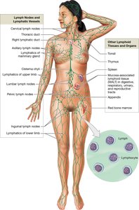
The lymphatic system is a vital part of the body's defense mechanisms, responsible for the production, maintenance, and distribution of lymphocytes, as well as the return of fluids to the blood and the distribution of nutrients and waste.
Lymph: A fluid similar to plasma, transported by lymphatic vessels from tissues to veins.
Lymphatic vessels: Network that carries lymph throughout the body.
Lymphoid tissues and organs: Structures containing large numbers of lymphocytes, including primary organs (formation and maturation of lymphocytes) and secondary organs (activation and cloning of lymphocytes).
Primary lymphoid organs: Red bone marrow and thymus (site of lymphocyte formation and maturation).
Secondary lymphoid organs: Tonsils, spleen, appendix, and MALT (mucosa-associated lymphoid tissue).
Immunity Overview
Immunity is the ability to resist infection or disease. It is divided into two main types:
Innate (nonspecific) immunity: Present at birth, does not distinguish between threats.
Adaptive (specific) immunity: Develops after exposure to specific antigens, involves T and B cells.
Innate Immunity
Physical and Chemical Barriers
Innate immunity includes physical barriers and chemical defenses that prevent pathogens from entering th+6 body.
Physical barriers: Skin, mucus, urine, stomach acid.
Chemical barriers: Sebaceous glands secrete chemicals, lysozymes, and antibodies.
Cellular Defenses
Phagocytes: Remove cellular debris and respond to invasion. Includes microphages (neutrophils, eosinophils) and macrophages (derived from monocytes).
Natural killer (NK) cells: Detect and destroy abnormal cells, including infected and cancer cells.
Chemical Defenses
Interferons: Released by lymphocytes, macrophages, and infected cells; interfere with viral replication.
Complement system: Chain reaction of proteins that attract phagocytes, destroy plasma membranes, and promote inflammation.
Inflammation and Fever
Mast cells: Release heparin and histamine, increasing blood flow and vessel permeability, causing swelling, redness, heat, and pain.
Fever: Pyrogens raise body temperature, increasing metabolism and inhibiting some bacteria.
Adaptive Immunity
Types of Adaptive Immunity
Adaptive immunity responds to specific threats and involves T and B cells.
Active immunity: Develops after exposure to an antigen (natural or artificial, e.g., vaccines).
Passive immunity: Results from the transfer of antibodies (natural, e.g., placenta or breastmilk; artificial, e.g., injected antibodies).
Characteristics of Adaptive Immunity
Specificity: Responses are specific to each antigen.
Versatility: Large number of lymphocytes ready to respond.
Memory: Memory cells remember previously encountered antigens.
Tolerance: Normal and self tissues are generally ignored.
Lymphocytes
T Cells
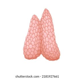
T cells provide cell-mediated immunity and mature in the thymus.
Cytotoxic T cells: Directly attack foreign or infected cells.
Helper T cells: Stimulate T and B cells.
Regulatory T cells: Inhibit T and B cells.
Memory T cells: Respond to previously encountered antigens.
B Cells
B cells provide humoral immunity (antibody-mediated immunity) and mature in the bone marrow.
Plasma cells: Produce antibodies.
Memory B cells: Respond to future exposures.
Natural Killer Cells
NK cells provide innate immunity by attacking foreign, infected, and cancer cells.
Lymphocyte Formation
Hematopoietic stem cells in red bone marrow produce lymphoid stem cells.
Some stay in bone marrow to make B and NK cells.
Some migrate to the thymus to make T cells.
B and T cells undergo positive (does the cell work?) and negative (does the cell react to foreign substances?) selection.
T Cell Activation and Function
Antigen Presentation and Recognition
T cells are activated by exposure to antigens bound to major histocompatibility complex (MHC) proteins.
Class I MHC: Found in all nucleated cells; recognized by CD8 T cells.
Class II MHC: Found in lymphocytes and antigen-presenting cells; recognized by CD4 T cells.
Activation and Cell Division
After binding to the MHC-antigen complex, costimulation occurs, preventing self-tissue attack. Activated T cells divide and differentiate.
Destruction of Target Cells
Cytotoxic T cells (CD8) attack infected cells using:
Perforin: Ruptures plasma membrane.
Cytokines: Activate apoptosis.
Lymphotoxin: Disrupts cell metabolism.
Memory cells ensure a faster and stronger response upon later exposures. 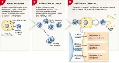
B Cell Activation and Antibody Production
Sensitization and Activation
B cells are sensitized when antigens enter via endocytosis and are displayed on Class II MHC proteins. Helper T cells bind and secrete cytokines, promoting B cell activation and division.
Cell Division and Differentiation
Activated B cells differentiate into plasma cells (produce antibodies) and memory B cells (respond to future exposures). 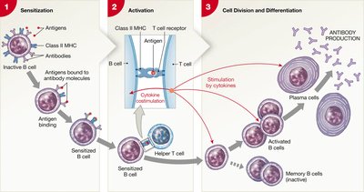
Antibody Structure and Function
Antibodies are Y-shaped molecules with constant segments forming the base and variable segments forming the antigen-binding site.
Neutralization: Prevents bacteria from binding to cells.
Agglutination: Clumps antigens for removal.
Activation of complement: Enhances phagocytosis and inflammation.
Opsonization: Makes pathogens easier to engulf.
Stimulation of inflammation: Promotes immune response.
Classes of Antibodies (Immunoglobulins)
IgG: Largest and most diverse; crosses placenta.
IgM: First antibody secreted after exposure; found on blood cells.
IgA: Found in mucus, tears, and saliva.
IgE: Stimulates basophils and mast cells to trigger inflammation.
IgD: On the surface of B cells.
Immune Responses
Primary and Secondary Responses
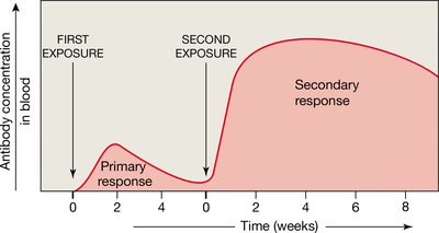
Primary response: Initial response to an antigen; slower, antibody levels peak after 1-2 weeks.
Secondary response: Memory cells differentiate immediately, causing a faster and stronger response.
Abnormal Immune Responses
Autoimmune Disorders
B cells make antibodies against normal cells, leading to diseases such as rheumatoid arthritis, Type 1 diabetes, and psoriasis.
Immunodeficiency
The immune system fails to develop or is blocked, as seen in SCID (failure to develop cell or antibody-mediated immunity) and AIDS (HIV destroys helper and regulatory T cells).
Allergies and Organ Rejection
Allergies: Excessive immune responses to non-harmful antigens; may destroy normal cells.
Anaphylaxis: Mast cells release histamine, causing severe reactions.
Organ rejection: T cells are activated by non-self MHC proteins; immunosuppressive drugs can decrease rejection risk but may compromise immunity.
Review Questions
What type of MHC protein do cytotoxic T cells recognize?
What do helper T cells do?
How are B cells sensitized?
Why would immunosuppressive drugs be helpful? When would they not?
Additional info: These notes expand brief lecture points into full academic explanations, including definitions, examples, and logical grouping for clarity.
