 Indietro
The Lymphatic System and Lymphoid Organs & Tissues: Study Guide
Guida di studio - Note intelligenti
 Appunti personalizzati basati sui tuoi materiali, ampliati con definizioni chiave, esempi e contesto.
The Lymphatic System
Function of the Lymphatic System
The lymphatic system is essential for maintaining fluid balance in the body and supporting immune function. It returns interstitial fluid and leaked plasma proteins back to the blood, preventing edema and ensuring proper circulation.
Returns interstitial fluid: Approximately 3 liters per day are returned to the bloodstream via lymphatic vessels.
Lymphatics: Network of lymphatic vessels that transport lymph.
Lymph: Interstitial fluid once it enters lymphatic vessels.
Lymph nodes: Act as filters, cleansing lymph before it re-enters the blood.
Distribution and Structure of Lymphatics
Lymphatic Capillaries
Lymphatic capillaries are highly permeable, blind-ended vessels that absorb interstitial fluid and larger molecules.
Absent from: Bones, teeth, bone marrow, and central nervous system (CNS).
Permeability: Allows uptake of proteins, pathogens, and cancer cells.
One-way mini-valves: Ensure fluid enters but does not exit capillaries.
Collagen anchors: Secure mini-valves to the matrix.
Lacteals: Specialized capillaries in intestinal mucosa that absorb digested fat and deliver fatty lymph (chyle) to the blood.
Collecting Lymphatic Vessels, Trunks, and Ducts
Lymphatic capillaries drain into collecting vessels, which then form trunks and ducts.
Collecting vessels: Similar to veins but with thinner walls and more valves.
Lymphatic trunks: Named for the regions they drain.
Lymphatic ducts: Empty lymph into venous circulation at the junction of internal jugular and subclavian veins.
Right lymphatic duct: Drains right upper arm, right head, and thorax.
Thoracic duct: Drains the rest of the body; begins at the cisterna chyli.
Lymphangitis
Lymphangitis is inflammation of the vasa vasorum supplying larger lymphatic vessels.
Causes: Infection or inflammation.
Treatment: Antibiotics and anti-inflammatory drugs.
Lymph Transport
Lymph is transported through a low-pressure system propelled by several mechanisms.
Skeletal muscle pump
Respiratory pump
Valves: Numerous, prevent backflow.
Pulsation of nearby arteries
Contraction of smooth muscle in lymphatics
Lymphedema
Lymphedema occurs when lymph cannot return to the blood, causing swelling.
Causes: Tumors blocking lymphatics, removal of lymphatics.
Treatment: Exercise, massage, compression.
Lymphoid Cells
Immune System Cells
Lymphoid cells are crucial for immune defense.
Lymphocytes: Originate in bone marrow and mature into T and B cells.
T cells (T lymphocytes): Manage immune response and destroy infected cells.
B cells (B lymphocytes): Produce plasma cells that secrete antibodies.
Macrophages: Phagocytize invaders and activate T cells.
Dendritic cells: Capture antigens and deliver them to lymph nodes; activate T cells.
Supporting Lymphoid Cells
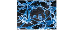
Reticular cells produce reticular fibers, forming the stroma—a network-like scaffolding for immune cells.
Stroma: Provides structural support for lymphocytes and macrophages.
Lymphoid Tissue
Structure and Function
Lymphoid tissue is composed of reticular connective tissue, providing a site for immune cell proliferation and surveillance.
Macrophages: Reside on reticular fibers.
Lymphocytes: Occupy spaces between fibers.
Function: Houses lymphocytes and offers vantage points for immune surveillance.
Types of Lymphoid Tissue
Diffuse lymphoid tissue: Found in almost every organ, especially in lamina propria.
Lymphoid follicles (nodules): Tightly packed lymphoid cells and fibers, contain germinal centers of proliferating B cells. Peyer’s patches and appendix are examples of isolated aggregations.
Lymphoid Organs
Classification
Primary lymphoid organs: Sites where T and B cells mature (e.g., bone marrow, thymus).
Secondary lymphoid organs: Sites where mature lymphocytes encounter antigens and become activated (e.g., lymph nodes, spleen, MALT).
Lymph Nodes
Function
Lymph nodes filter lymph and activate the immune system.
Cleanse lymph: Macrophages destroy microorganisms and debris.
Activate immune system: Lymphocytes become activated upon encountering antigens.
Structure
Cortex: Contains B cells in germinal centers, T cells in deep cortex, and dendritic cells.
Medulla: Contains B cells, T cells, and plasma cells.
Sinuses: Large lymphatic capillaries and reticular fibers with macrophages.
External fibrous capsule: Extends inward as trabeculae.
Circulation in Lymph Nodes
Lymph enters via afferent vessels, passes through subscapular and medullary sinuses, and exits at the hilum via efferent vessels.
Buboes
Buboes are inflamed, swollen, and tender lymph nodes overwhelmed by pathogens.
Spleen
Functions
The spleen is the largest lymphoid organ, involved in immune response and blood filtration.
Lymphocyte proliferation and immune response
Cleanses blood: Removes aged blood cells and platelets.
Stores breakdown products of RBCs: e.g., iron.
Stores platelets and monocytes
Site of fetal erythropoiesis
Structure
Fibrous capsule and trabeculae
White pulp: Immune function, contains lymphocytes on reticular fibers.
Red pulp: Destruction of old blood cells and pathogens; composed of splenic cords and venous sinuses.
Splenectomy
Causes: Rupture due to trauma or infection.
After removal: Liver and bone marrow compensate for most functions.
Regeneration: Possible in children under 12 years.
MALT (Mucosa-Associated Lymphoid Tissue)
Function and Location
MALT protects mucosal surfaces from pathogens.
Location: Mucous membranes of respiratory, genitourinary, and gastrointestinal tracts.
Largest collections: Tonsils, Peyer’s patches, appendix.
Tonsils
Function and Structure
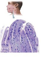
Tonsils gather and remove pathogens from food and air.
Location: Named according to anatomical position (pharyngeal, palatine, lingual).
Structure: Partial capsule, follicles with germinal centers and lymphocytes.
Tonsillar crypts: Invaginations in the epithelium that trap pathogens.
Peyer’s Patches
Function and Location
Peyer’s patches are clusters of lymphoid follicles in the ileum of the small intestine.
Function: Destroy bacteria and generate memory lymphocytes.
Appendix
Function and Structure
The appendix is an offshoot of the large intestine, containing lymphoid tissue.
Function: Destroy bacteria and generate memory lymphocytes.
Appendicitis
Causes: Blockage by stool, foreign bodies, or cancer.
Treatment: Antibiotics, pus removal, appendectomy if ruptured.
Thymus
Function and Structure
The thymus is a bi-lobed organ in the inferior neck, crucial for T cell maturation.
Large during childhood: Gradually atrophies with age.
No follicles: No B cells present.
Does not directly fight antigens: Blood-thymus barrier isolates immature T cells.
Stroma: Made of epithelial cells, not reticular fibers.
Structure
Cortex: Contains lymphocytes.
Medulla: Contains Hassall’s corpuscles, where regulatory T cells develop (prevent autoimmunity).
