 Indietro
The Lymphatic System and Lymphoid Organs & Tissues: Study Guide
Guida di studio - Note intelligenti
 Appunti personalizzati basati sui tuoi materiali, ampliati con definizioni chiave, esempi e contesto.
The Lymphatic System: Overview
Major Functions of the Lymphatic System
The lymphatic system is a crucial component of the circulatory and immune systems, responsible for returning fluids leaked from blood vessels back to the blood, and providing structural support for immune responses.
Fluid Recovery: Returns excess interstitial fluid (about 3L/day) to the bloodstream.
Immune Defense: Houses lymphoid organs and tissues that support immune cell activation and proliferation.
Absorption of Fats: Specialized lymphatic capillaries (lacteals) absorb fats from the intestine.
Components of the Lymphatic System
Lymphatic Vessels: Network of drainage vessels that transport lymph.
Lymph: Fluid within lymphatic vessels, similar to plasma but with less protein.
Lymph Nodes: Structures that filter lymph and activate immune responses.
Lymphoid Organs: Include spleen, thymus, tonsils, lymph nodes, and other tissues.
Lymphatic Circulation and Vessel Structure
Lymphatic Vessels and Capillaries
Lymphatic vessels begin as blind-ended capillaries in tissues, collecting interstitial fluid and returning it to the blood. They are more permeable than blood capillaries, allowing uptake of proteins, pathogens, and cell debris.
Lymphatic Capillaries: Loosely overlapping endothelial cells form one-way minivalves, anchored by collagen filaments. Increased interstitial fluid volume opens minivalves, allowing fluid entry.
Glymphatic System: In the brain, astrocytes form channels connecting to lymphatics in the meninges, aiding in waste drainage.
Lacteals: Specialized capillaries in the small intestine absorb digested fats, forming chyle (fatty lymph).
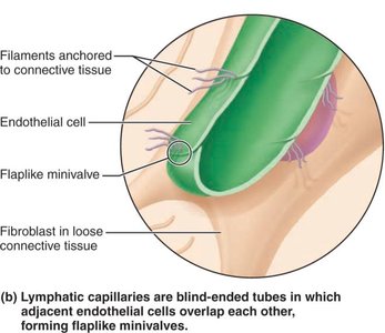
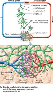
Example: In inflamed tissues, lymphatic capillaries can take up larger substances, including pathogens and cancer cells.
Major Lymphatic Trunks and Ducts
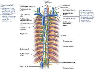
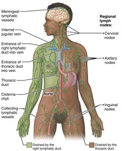
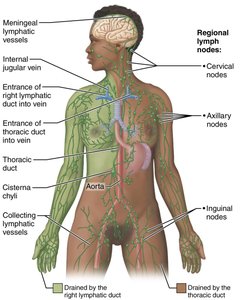
Lymphatic vessels converge to form trunks and ducts, which return lymph to the venous system at the junctions of the internal jugular and subclavian veins.
Right Lymphatic Duct: Drains lymph from the right upper limb, right side of head and thorax.
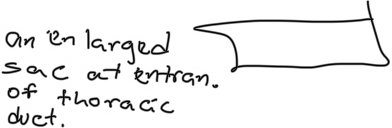
Thoracic Duct: Drains lymph from the rest of the body; begins at the cisterna chyli, an enlarged sac at the entrance.
Lymph Transport Mechanisms
How Lymph Moves
Lymphatic vessels are low-pressure conduits, relying on mechanisms similar to veins for unidirectional flow.
Milking Action: Skeletal muscle contractions push lymph forward.
Respiratory Movements: Pressure changes during breathing aid lymph flow.
Arterial Pulsations: Pulse pressure waves from adjacent arteries help move lymph.
Smooth Muscle Contractions: Rhythmic contractions in vessel walls (except smallest lymphatics).
Clinical Note: Physical activity increases lymph flow; immobilization of infected areas restricts spread of inflammatory materials.
Lymphoid Tissue and Organs
Lymphoid Tissue
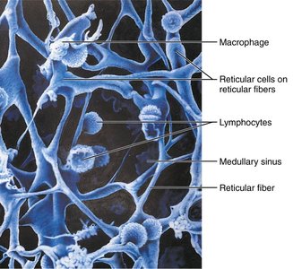
Lymphoid tissue provides sites for lymphocyte activation and proliferation, and is an ideal surveillance location for immune cells.
Reticular Connective Tissue: Most lymphoid tissues are composed of this tissue, except the thymus, which is made of epithelium.
Primary and Secondary Lymphoid Organs
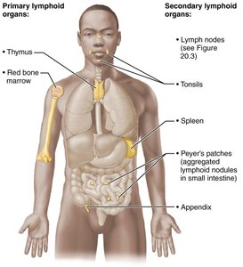
Lymphoid organs are categorized based on their function in immune cell development and activation.
Primary Lymphoid Organs: Sites where T and B cells mature (red bone marrow and thymus).
Secondary Lymphoid Organs: Sites where mature lymphocytes encounter antigens and become activated (lymph nodes, spleen, tonsils, Peyer's patches, appendix).
Lymph Nodes: Structure and Function
Anatomy and Protective Functions
Lymph nodes are distributed throughout the body, with large clusters near the inguinal, axillary, and cervical regions. They serve two main protective functions:
Cleansing the Lymph: Act as filters, removing pathogens and debris.
Immune Activation: Sites where lymphocytes encounter antigens and mount immune responses.
Lymph Node Circulation
Lymph flows through nodes in a specific sequence:
Enters via afferent lymphatic vessels.
Travels through subcapsular sinus, then smaller sinuses in cortex and medulla.
Enters medullary sinuses.
Exits at hilum via efferent lymphatic vessels.
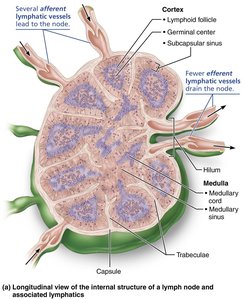
Clinical Relevance: Buboes and Cancer
Buboes: Infected, pus-filled lymph nodes (e.g., bubonic plague).
Lymphadenopathy: Swollen, tender nodes due to infection.
Cancer Metastasis: Cancer-infiltrated nodes are swollen but usually not painful.
The Spleen: Structure and Functions
Anatomy and Role
The spleen is the largest lymphoid organ, located in the upper left abdominal cavity. It provides a site for lymphocyte proliferation, immune surveillance, and response, and cleanses blood of aged cells and platelets.
Additional Functions: Stores iron, recycles breakdown products of red blood cells, stores platelets and monocytes, and may produce erythrocytes in the fetus.
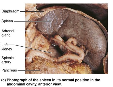
Spleen Histology: White and Red Pulp
White Pulp: Site of immune function, composed mostly of lymphocytes on reticular fibers around central arteries.
Red Pulp: Site of destruction of old blood cells and pathogens, rich in macrophages and RBCs.
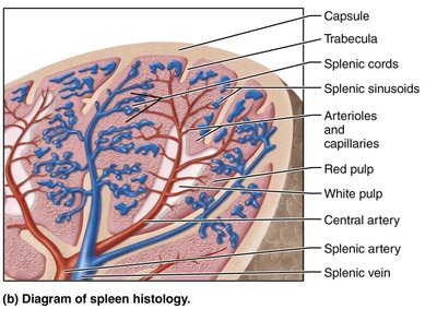
Mucosa-Associated Lymphoid Tissue (MALT)
Definition and Major Collections
MALT consists of lymphoid tissues in mucous membranes throughout the body, providing immune protection at entry points.
Tonsils: Located in the pharynx; trap and remove pathogens.
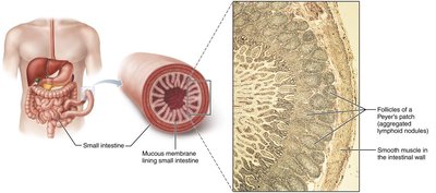
Peyer's Patches: Aggregated lymphoid nodules in the small intestine.
Appendix: Contains lymphoid tissue; may play a role in immunity.
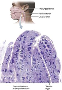
Summary Table: Lymphatic System Components
Component | Main Function | Location |
|---|---|---|
Lymphatic Vessels | Drain interstitial fluid, transport lymph | Throughout body |
Lymph Nodes | Filter lymph, activate immune response | Clusters in inguinal, axillary, cervical regions |
Spleen | Immune surveillance, cleanses blood | Upper left abdominal cavity |
Thymus | T cell maturation | Superior mediastinum |
Tonsils | Trap pathogens | Pharynx |
Peyer's Patches | Immune surveillance in intestine | Small intestine |
Appendix | Immune function | Large intestine |
Key Terms and Definitions
Lymph: Fluid collected by lymphatic vessels, similar to plasma.
Lymphatic Capillaries: Highly permeable vessels that collect interstitial fluid.
Lymph Node: Small, bean-shaped organ that filters lymph.
Lacteal: Specialized lymphatic capillary in the small intestine for fat absorption.
Chyle: Fatty lymph absorbed from the intestine.
MALT: Mucosa-associated lymphoid tissue.
Cisterna Chyli: Enlarged sac at the entrance of the thoracic duct.
Relevant Equations
There are no specific equations for lymphatic flow, but the following general relationship applies:
Fluid Movement: Driven by hydrostatic and osmotic pressure differences between blood capillaries and interstitial fluid.
Additional info: The lymphatic system is essential for maintaining fluid balance, immune defense, and fat absorption. Its dysfunction can lead to edema, immune deficiencies, or spread of cancer cells.
