 Indietro
The Lymphatic System and Lymphoid Organs: Structure, Function, and Clinical Significance
Guida di studio - Note intelligenti
 Appunti personalizzati basati sui tuoi materiali, ampliati con definizioni chiave, esempi e contesto.
The Lymphatic System and Lymphoid Organs
Overview of the Lymphatic System
The lymphatic system is a vital component of the circulatory and immune systems. It returns fluids leaked from blood vessels back to the blood, provides structural support for immune responses, and houses lymphoid organs and tissues that defend against pathogens.
Lymphatic vessels: Network of drainage vessels that collect excess interstitial fluid.
Lymph: Fluid within lymphatic vessels, derived from interstitial fluid.
Lymph nodes: Structures that cleanse lymph and house immune cells.
Lymphoid organs and tissues: Include spleen, thymus, tonsils, lymph nodes, and other tissues; provide the basis for immune defense.
Components of the Lymphatic System
Lymphatic Vessels
Lymphatic vessels collect excess protein-containing interstitial fluid and return it to the blood. They also transport pathogens to lymph nodes and absorbed fats from the intestine to the blood.
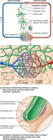
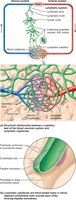
Lymphatic capillaries: Blind-ended vessels, highly permeable due to overlapping endothelial cells forming one-way minivalves and anchoring collagen filaments.
Glymphatic system: In the brain, astrocytes form channels connecting to lymphatics in the meninges, aiding in drainage of interstitial and cerebrospinal fluids.
Lacteals: Specialized lymphatic capillaries in the small intestine for absorption and transport of digested fat; fatty lymph (chyle) enters the blood.
Larger Lymphatic Vessels
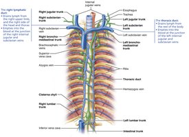
Lymphatic capillaries drain into larger collecting vessels, which have thinner walls, more valves, and more anastomoses than veins. These vessels unite to form nine major lymphatic trunks, named for the regions they drain.
Major trunks: Paired lumbar, bronchomediastinal, subclavian, jugular; single intestinal trunk.
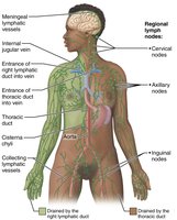
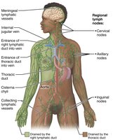
Lymph is delivered from trunks into two large ducts: Right lymphatic duct (drains right upper limb, head, thorax) and thoracic duct (drains rest of body).
Cisterna chyli: Enlarged sac at the base of the thoracic duct in about half of individuals.
Lymph Transport
Lymphatic vessels are low-pressure conduits, relying on mechanisms similar to veins for unidirectional flow:
Milking action of skeletal muscles
Pressure changes during breathing
Pulses from adjacent arteries
Rhythmic contractions of smooth muscle in vessel walls
One-way valves prevent backflow
Physical activity increases lymph flow; immobilization restricts outflow of inflammatory materials.
Clinical Significance
Lymphangitis: Inflammation of lymphatic vessels, appearing as red streaks.
Lymphedema: Severe localized edema due to insufficient lymph return, often caused by tumors or surgical removal of lymphatics.
Lymphoid Cells and Tissues
Lymphoid Cells
Lymphoid cells include immune system cells and supporting cells that form the structural framework of lymphoid tissues.
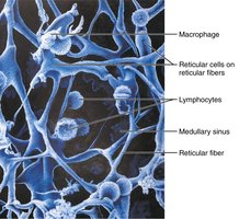
Lymphocytes: T cells (manage immune response, attack infected cells) and B cells (produce plasma cells, secrete antibodies).
Macrophages: Phagocytize foreign substances and activate T cells.
Dendritic cells: Capture and deliver antigens to lymph nodes.
Reticular cells: Produce reticular fiber stroma, supporting other cell types.
Lymphoid Tissue
Lymphoid tissue is a key component of the immune system, providing sites for activation and proliferation of lymphocytes and macrophages. It is largely composed of loose reticular connective tissue.
Diffuse lymphoid tissue: Loose arrangement of cells and fibers, found in most organs.
Lymphoid follicles (nodules): Solid, spherical bodies with germinal centers of proliferating B cells; found in lymph nodes, Peyer's patches, and appendix.
Lymphoid Organs
Primary and Secondary Lymphoid Organs
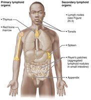
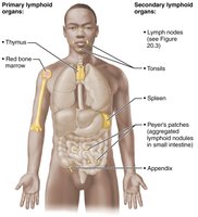
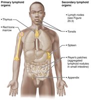
Lymphoid organs are classified as primary (where lymphocytes mature) or secondary (where mature lymphocytes encounter antigens).
Primary lymphoid organs: Red bone marrow (B cell maturation), thymus (T cell maturation).
Secondary lymphoid organs: Lymph nodes, spleen, tonsils, Peyer's patches, appendix.
Lymph Nodes
Structure and Function
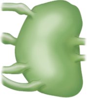
Lymph nodes are bean-shaped, small structures clustered along lymphatic vessels. They cleanse lymph and house lymphocytes.
Cleansing: Macrophages remove microorganisms and debris.
Immune activation: Lymphocytes encounter antigens and mount immune responses.
Located in clusters near body surface (inguinal, axillary, cervical regions).
Internal Structure
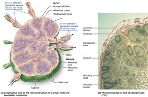
Capsule: External fibrous covering.
Trabeculae: Fibers extending inward, dividing node into compartments.
Cortex: Contains follicles with germinal centers (B cells), T cells, dendritic cells.
Medulla: Medullary cords with B cells, T cells, plasma cells.
Lymph sinuses: Capillaries spanned by reticular fibers; macrophages reside here.
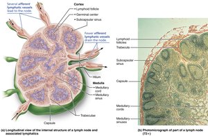
Circulation
Lymph enters via afferent vessels, passes through sinuses, exits via efferent vessels at the hilum.
Fewer efferent vessels than afferent, slowing lymph flow and allowing time for immune functions.
Clinical Significance
Buboes: Infected, pus-filled lymph nodes (e.g., bubonic plague).
Lymphadenopathy: Swollen, tender nodes due to infection.
Cancer metastasis: Cancer cells may become trapped in lymph nodes, causing painless swelling.
The Spleen
Structure and Function
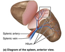
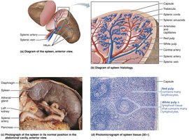
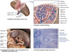
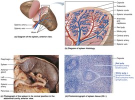
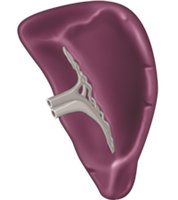
The spleen is the largest lymphoid organ, located in the left abdominal cavity. It provides sites for lymphocyte proliferation, immune surveillance, and response, and cleanses blood of aged cells and platelets.
Stores iron and recycles breakdown products of red blood cells.
Stores blood platelets and monocytes.
May produce erythrocytes in the fetus.
Histology
White pulp: Immune function; lymphocytes on reticular fibers around central arteries.
Red pulp: Destruction of old blood cells and pathogens; contains RBCs and macrophages.
Clinical Significance
Splenic rupture: May require splenectomy; liver and bone marrow compensate for lost functions.
Children's spleen can regenerate if a portion remains.
Mucosa-Associated Lymphoid Tissue (MALT)
Structure and Function
MALT consists of lymphoid tissues in mucous membranes throughout the body, protecting against pathogens entering via respiratory, genitourinary, and digestive tracts.
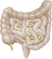
Largest collections: Tonsils, Peyer's patches, appendix.
Tonsils
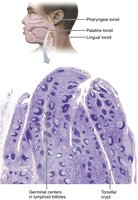
Form a ring of lymphoid tissue around the entrance to the pharynx.
Gather and remove pathogens from food and air.
Contain follicles with germinal centers and tonsillar crypts for trapping debris.
Peyer's Patches
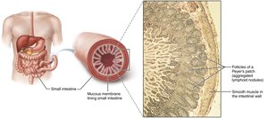
Large clusters of lymphoid follicles in the wall of the distal small intestine.
Prevent bacteria from breaching the intestinal wall and generate memory lymphocytes.
Appendix
Tubular offshoot of the large intestine containing lymphoid follicles.
Functions similar to Peyer's patches.
Thymus
Structure and Function
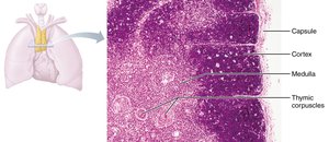
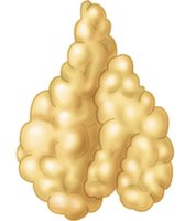
The thymus is a bilobed lymphoid organ in the inferior neck and mediastinum, prominent in childhood. It is the site of T lymphocyte development and maturation.
Divided into lobules with cortex (rapidly dividing lymphocytes, macrophages) and medulla (fewer lymphocytes, thymic corpuscles).
Thymic corpuscles involved in regulatory T cell development.
Differs from secondary lymphoid organs: lacks follicles, does not directly fight antigens, blood-thymus barrier prevents premature activation.
Stroma consists of epithelial cells, not reticular fibers.
Summary Table: Lymphoid Organs and Tissues
The following table summarizes the main lymphoid organs and tissues, their structure, and function.
Organ/Tissue | Structure | Function |
|---|---|---|
Lymph Node | Bean-shaped, cortex and medulla, follicles | Filters lymph, immune activation |
Spleen | White pulp, red pulp, fibrous capsule | Filters blood, immune response, stores platelets |
Tonsils | Follicles, crypts | Trap pathogens, immune memory |
Peyer's Patches | Follicles in small intestine | Prevent bacterial invasion, immune memory |
Appendix | Follicles in large intestine | Prevent bacterial invasion, immune memory |
Thymus | Lobules, cortex, medulla, corpuscles | T cell maturation |
Developmental Aspects
Lymphatic vessels and main clusters of lymph nodes develop by the fifth week of embryonic development. Lymphoid organs (except thymus) arise from mesodermal mesenchymal cells. The thymus is an endodermal derivative and the first lymphoid organ to appear.
Thymus develops from primitive pharynx lining.
Other lymphoid organs become populated by lymphocytes shortly after birth.
Key Terms and Concepts
Lymph: Interstitial fluid that enters lymphatic vessels.
Antigen: Substance that triggers an immune response.
Chyle: Fatty lymph absorbed from the intestine.
Memory lymphocytes: Cells that provide long-term immunity.
Equations and Formulas
Fluid movement across capillaries is governed by hydrostatic and osmotic pressures:
Net filtration pressure (NFP):
This equation explains the movement of fluid into and out of capillaries, contributing to lymph formation.
Additional info:
Red bone marrow, a primary lymphoid organ, is not included in some summary tables.
MALT includes diffuse lymphoid tissues in mucosa-associated regions.
