Indietro
The Lymphatic System: Structure, Function, and Immune Role
Guida di studio - Note intelligenti
 Appunti personalizzati basati sui tuoi materiali, ampliati con definizioni chiave, esempi e contesto.
The Lymphatic System: Overview and Functions
Introduction to the Lymphatic System
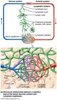
The lymphatic system is a crucial component of the circulatory and immune systems. It returns interstitial fluid and leaked plasma proteins back to the blood, provides a route for fat absorption, and houses immune cells for body defense.
Lymphatic vessels form a one-way system, ensuring lymph flows only toward the heart.
Major structures include lymphatic capillaries, collecting lymphatic vessels, lymphatic trunks, and lymphatic ducts.
Lymph is eventually returned to venous circulation, maintaining fluid balance.
Functions of Lymphatic Vessels
Return excess tissue fluid to the blood.
Return leaked proteins to the blood.
Carry pathogens to lymph nodes for immune surveillance.
Transport absorbed fats from the intestine to the blood via specialized lymphatic capillaries called lacteals.
Structure and Distribution of Lymphatic Vessels
Lymphatic Vessel Organization
Lymphatic vessels are distributed throughout the body and are organized into a hierarchical system:
Lymphatic capillaries (smallest, highly permeable)
Collecting lymphatic vessels
Lymphatic trunks (drain large body regions)
Lymphatic ducts (return lymph to venous circulation)
Lymphatic Trunks and Ducts
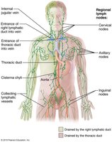
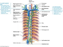
Lymphatic trunks are named for the regions they drain: paired lumbar, bronchomediastinal, subclavian, jugular trunks, and a single intestinal trunk.
Lymph is delivered from trunks into two main ducts:
Right lymphatic duct: drains right upper arm and right side of head and thorax.
Thoracic duct: drains the rest of the body; often begins as an enlarged sac called the cisterna chyli.
Each duct empties lymph into venous circulation at the junction of the internal jugular and subclavian veins.
Lymphoid Cells, Tissues, and Organs
Lymphoid Cells
Lymphoid cells are essential for immune function and include both immune system cells and supporting cells.
Lymphocytes: Main cells of the adaptive immune system; two types:
T cells (T lymphocytes): Mediate cellular immunity.
B cells (B lymphocytes): Produce antibodies for humoral immunity.
Other immune cells: Macrophages (phagocytosis), dendritic cells (antigen presentation).
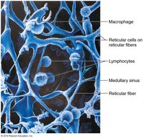
Supporting cells: Reticular cells produce reticular fibers (stroma) that form the structural network of lymphoid organs.
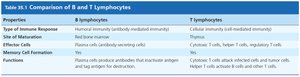
Comparison of B and T Lymphocytes
Properties | B Lymphocytes | T Lymphocytes |
|---|---|---|
Type of Immune Response | Humoral (antibody-mediated) | Cellular (cell-mediated) |
Site of Maturation | Red bone marrow | Thymus |
Effector Cells | Plasma cells (antibody-secreting) | Cytotoxic T cells, helper T cells, regulatory T cells |
Memory Cell Formation | Yes | Yes |
Functions | Produce antibodies that inactivate antigens and tag them for destruction | Attack infected and tumor cells; activate other immune cells |
Lymphoid Tissue
Lymphoid tissue is primarily reticular connective tissue that houses and provides activation sites for lymphocytes and macrophages. It offers surveillance vantage points for immune cells.
Lymphoid Organs
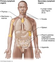
Classification of Lymphoid Organs
Lymphoid organs are divided into primary and secondary categories based on their function in lymphocyte development and activation.
Primary lymphoid organs: Sites where lymphocytes mature.
Red bone marrow: Origin and maturation site for B cells.
Thymus: Maturation site for T cells.
Secondary lymphoid organs: Sites where mature lymphocytes encounter antigens and become activated.
Includes lymph nodes, spleen, MALT (mucosa-associated lymphoid tissue), Peyer’s patches, appendix, and diffuse lymphoid tissues.
Lymph Nodes
Structure and Function of Lymph Nodes
Lymph nodes are the principal secondary lymphoid organs, distributed throughout the body, often in clusters along lymphatic vessels. They are especially concentrated in the inguinal, axillary, and cervical regions.
Cleansing the lymph: Act as filters, removing unwanted substances before lymph is returned to the blood.
Immune system activation: Provide sites for lymphocyte activation and mounting of immune responses.
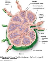
Internal Structure of a Lymph Node
Bean-shaped, less than 2.5 cm in size.
Surrounded by a fibrous capsule; capsule fibers extend inward as trabeculae, dividing the node into compartments.
Two main regions:
Cortex: Contains follicles with germinal centers (B cells) and deeper areas with T cells.
Medulla: Contains medullary cords (B cells, T cells, plasma cells) and lymph sinuses (large capillaries with reticular fibers and macrophages).
Circulation in the Lymph Nodes
Lymph enters via afferent lymphatic vessels on the convex side.
Flows through subcapsular sinus, then smaller sinuses in cortex and medulla, and finally exits at the hilum via efferent vessels.
Fewer efferent vessels slow lymph flow, allowing immune cells time to act.
Lymph typically passes through several nodes before returning to circulation.
MALT: Mucosa-Associated Lymphoid Tissue
Definition and Locations
MALT consists of lymphoid tissues located in mucous membranes throughout the body, providing protection against pathogens entering through mucosal surfaces.
Found in the mucosa of the respiratory tract, genitourinary organs, and digestive tract.
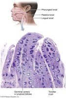
Largest collections are in the tonsils, Peyer’s patches (small intestine), and appendix.