 Indietro
The Lymphatic System: Structure, Function, and Clinical Relevance
Guida di studio - Note intelligenti
 Appunti personalizzati basati sui tuoi materiali, ampliati con definizioni chiave, esempi e contesto.
The Lymphatic System
Overview of the Lymphatic System
The lymphatic system is a crucial component of the circulatory and immune systems. It is responsible for returning interstitial fluid to the bloodstream, absorbing dietary fats, and providing immune defense. The lymphatic system consists of lymphatic vessels, lymph nodes, and lymphoid organs.
The Cardiovascular System and Its Relationship to the Lymphatic System
Pathway of Blood Through the Heart and Body
The cardiovascular system works closely with the lymphatic system to maintain fluid balance and transport nutrients and waste. Blood flows through the heart in a specific sequence, passing through chambers and valves before circulating to the lungs and body tissues.
Right Atrium → Tricuspid Valve → Right Ventricle → Pulmonary Semilunar Valve → Pulmonary Trunk → Lungs → Pulmonary Veins → Left Atrium → Bicuspid Valve → Left Ventricle → Aortic Semilunar Valve → Aorta → Systemic Capillaries → Vena Cava (back to right atrium)
Oxygenated blood is shown in red; deoxygenated blood is shown in blue.
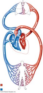

Systemic and Pulmonary Circuits
The heart pumps blood through two main circuits:
Pulmonary Circuit: Carries deoxygenated blood from the right ventricle to the lungs and returns oxygenated blood to the left atrium.
Systemic Circuit: Delivers oxygenated blood from the left ventricle to the body and returns deoxygenated blood to the right atrium.
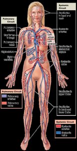
Structural Relationship Between Blood Capillaries and Lymphatic Capillaries
Capillary Beds and Fluid Exchange
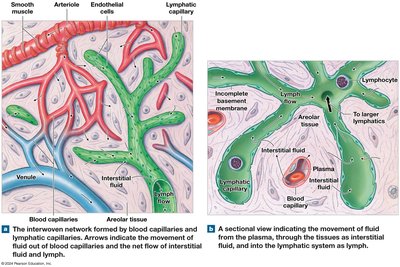
Blood capillaries and lymphatic capillaries are closely associated in tissues. Fluid and small solutes move out of blood capillaries into the interstitial space, where some is taken up by lymphatic capillaries to become lymph.
Blood Capillaries: Exchange nutrients, gases, and wastes with tissues.
Lymphatic Capillaries: Absorb excess interstitial fluid and return it to the bloodstream.
Interstitial Fluid: The fluid that surrounds tissue cells, derived from plasma.
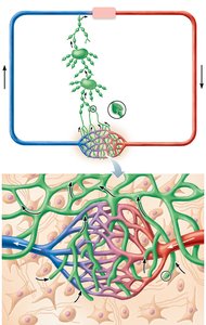
Movement of Fluid
Fluid moves from blood capillaries into tissues, forming interstitial fluid. Lymphatic capillaries collect this fluid, now called lymph, and transport it through lymphatic vessels back to the venous system.
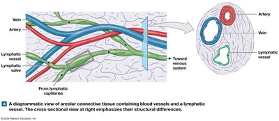
Comparison of Arteries, Veins, and Lymphatic Vessels
Structural Differences
Arteries, veins, and lymphatic vessels have distinct structural features:
Arteries: Thick walls, carry blood away from the heart under high pressure.
Veins: Thinner walls, carry blood toward the heart, often contain valves to prevent backflow.
Lymphatic Vessels: Thin walls, contain numerous valves, transport lymph toward the heart.
Major Lymphatic Vessels and Nodes
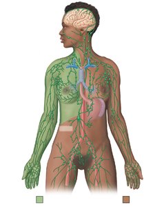
Distribution of Lymphatic Vessels and Regional Lymph Nodes
Lymphatic vessels collect lymph from tissues and transport it through lymph nodes, where immune responses can be initiated. Major lymphatic ducts include the thoracic duct and right lymphatic duct.
Thoracic Duct: Drains lymph from most of the body into the left subclavian vein.
Right Lymphatic Duct: Drains lymph from the right upper limb, right side of thorax, and right side of head and neck into the right subclavian vein.
Regional Lymph Nodes: Cervical, axillary, and inguinal nodes filter lymph from specific body regions.
Histology of Lymphoid Tissue
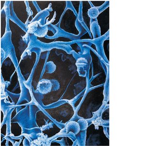
Reticular Connective Tissue
Lymphoid organs are supported by a network of reticular fibers, which provide a scaffold for immune cells such as lymphocytes and macrophages.
Reticular Connective Tissue: Contains reticular fibers and cells, forming the structural framework of lymphoid organs.
Macrophages: Phagocytic cells that engulf pathogens and debris.
Lymphocytes: Key cells of the adaptive immune response.
Lymphoid Organs
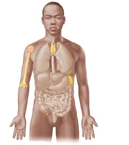
Primary and Secondary Lymphoid Organs
Lymphoid organs are classified as primary or secondary based on their roles in lymphocyte development and immune response.
Primary Lymphoid Organs | Secondary Lymphoid Organs |
|---|---|
Thymus | Lymph nodes |
Red bone marrow | Tonsils |
Spleen | |
Peyer’s patches | |
Appendix |
Primary lymphoid organs are where lymphocytes mature (thymus for T cells, red bone marrow for B cells). Secondary lymphoid organs are sites where immune responses are initiated.
Structure of a Lymph Node
Internal Anatomy of a Lymph Node
Lymph nodes filter lymph and house immune cells. They have a distinct structure with several key regions:
Afferent Lymphatic Vessels: Bring lymph into the node.
Capsule: Outer connective tissue covering.
Trabeculae: Extensions of the capsule into the node.
Cortex: Contains lymphoid follicles with germinal centers (B cell proliferation).
Paracortex: Rich in T cells.
Medulla: Contains medullary cords (B cells, macrophages) and sinuses.
Efferent Lymphatic Vessel: Drains filtered lymph from the node.
Specialized Lymphoid Tissues
Tonsils
Tonsils are lymphoid tissues located in the pharynx. They contain lymphoid follicles and crypts that trap pathogens.
Palatine Tonsil: Covered by non-keratinized stratified squamous epithelium, contains tonsillar crypts and lymphoid follicles with germinal centers.
Pharyngeal and Lingual Tonsils: Other tonsillar tissues with similar immune functions.
Peyer’s Patches and Appendix
Peyer’s patches are clusters of lymphoid follicles in the small intestine, while the appendix contains abundant lymphoid tissue. Both are examples of mucosa-associated lymphatic tissue (MALT).
Peyer’s Patches: Located in the ileum, monitor intestinal bacteria and prevent the growth of pathogenic bacteria.
Appendix: Contains lymphoid follicles, contributes to gut immunity.
Spleen
The spleen filters blood, removes old erythrocytes, and mounts immune responses. It contains white pulp (lymphocytes) and red pulp (erythrocyte breakdown).
White Pulp: Lymphatic tissue surrounding central arteries.
Red Pulp: Contains splenic cords and sinusoids for filtering blood.
Thymus
The thymus is the site of T cell maturation. It is larger in children and shrinks with age, being replaced by adipose tissue in adults.
Cortex: Densely packed with immature T cells.
Medulla: Contains mature T cells and Hassall’s corpuscles.
Additional info: The lymphatic system is essential for immune surveillance, fluid balance, and fat absorption. Disorders of the lymphatic system can lead to lymphedema, infections, and immune deficiencies.
