 Indietro
The Muscular System: Mechanisms of Skeletal Muscle Contraction
Guida di studio - Note intelligenti
 Appunti personalizzati basati sui tuoi materiali, ampliati con definizioni chiave, esempi e contesto.
The Muscular System
Nerve-Muscle Relationship
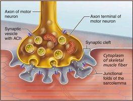
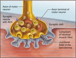
The interaction between nerves and muscles is fundamental for voluntary movement. Skeletal muscles are stimulated by somatic motor neurons, whose axons extend from the central nervous system to the muscle fibers. Each muscle fiber is innervated by a single motor neuron at a specialized synapse called the neuromuscular junction (NMJ).
Motor Neuron: A nerve cell that transmits signals to muscle fibers, causing contraction.
Neuromuscular Junction: The site where the axon terminal of a motor neuron meets the muscle fiber.
Synaptic Cleft: The gap between the axon terminal and the muscle fiber membrane.
Acetylcholine (ACh): The neurotransmitter released from synaptic vesicles to stimulate muscle contraction.
Excitation: Events at the Neuromuscular Junction
Muscle excitation begins when the motor neuron releases ACh into the synaptic cleft. ACh binds to receptors on the sarcolemma (muscle cell membrane), triggering an action potential that spreads across the muscle fiber.
Action Potential: An electrical signal that travels along the sarcolemma.
Junctional Folds: Increase surface area for ACh receptors, enhancing signal transmission.
Excitation-Contraction Coupling
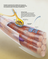
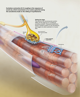
Excitation-contraction (E-C) coupling is the process by which the action potential leads to muscle contraction. The action potential travels along the sarcolemma and down T-tubules, causing voltage-sensitive proteins to stimulate the release of Ca2+ from the sarcoplasmic reticulum (SR). Calcium binds to troponin, exposing myosin-binding sites on actin and initiating contraction.
T-tubules: Invaginations of the sarcolemma that help transmit the action potential deep into the muscle fiber.
Sarcoplasmic Reticulum (SR): Organelle that stores and releases Ca2+.
Troponin: Protein that binds Ca2+ and moves tropomyosin away from actin's binding sites.
Sliding Filament Model of Contraction
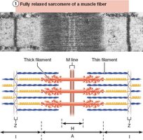
The sliding filament model explains how muscles contract. During contraction, thin filaments (actin) slide past thick filaments (myosin), increasing their overlap. Myosin heads bind to actin, forming cross bridges, and pull the actin filaments toward the center of the sarcomere.
Cross Bridge: The connection formed when myosin heads bind to actin.
Sarcomere: The functional unit of muscle contraction, defined by Z-discs.
Muscle Fiber Contraction: Cross Bridge Cycling
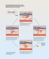
Cross bridge cycling is the sequence of events that causes muscle contraction. It requires Ca2+ to expose actin's binding sites. The cycle includes cross bridge formation, power stroke, detachment, and cocking of the myosin head.
Power Stroke: Myosin head pivots, pulling actin toward the M line.
Detachment: ATP binds to myosin, causing it to release actin.
Cocking: ATP hydrolysis re-energizes the myosin head.
Clinical Homeostatic Imbalance
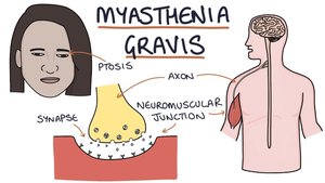
Disorders can disrupt neuromuscular function. Myasthenia gravis is an autoimmune disease where antibodies destroy ACh receptors, causing muscle weakness. Rigor mortis occurs after death when ATP is depleted, preventing cross bridge detachment and causing sustained contraction.
Myasthenia Gravis: Characterized by drooping eyelids, difficulty swallowing, and muscle weakness.
Rigor Mortis: Stiffening of muscles postmortem due to lack of ATP.
Whole Muscle Contraction
Muscle contraction produces tension, which is the force exerted by a muscle. The load is the opposing force. The force and duration of contraction depend on the frequency and intensity of stimuli.
Muscle Tension: Force generated by contracting muscle.
Load: The weight or resistance the muscle must overcome.
Isometric vs Isotonic Contractions
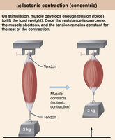
Muscle contractions can be classified as isometric or isotonic. Isometric contractions increase tension without shortening the muscle, while isotonic contractions involve muscle shortening and movement of a load. Isotonic contractions are further divided into concentric (muscle shortens) and eccentric (muscle lengthens).
Isometric Contraction: Muscle tension increases, but length remains unchanged.
Isotonic Contraction: Muscle changes length and moves a load.
Concentric: Muscle shortens and does work.
Eccentric: Muscle lengthens while generating force.
Muscle Metabolism
Muscle contraction requires ATP, which can be generated by anaerobic fermentation or aerobic respiration. Anaerobic fermentation converts glucose to lactate, producing 2 ATP per glucose without oxygen. Aerobic respiration uses oxygen to produce ~32 ATP per glucose, but is slower and cannot meet high demand during intense exercise.
Anaerobic Fermentation: Fast, low ATP yield, does not require oxygen.
Aerobic Respiration: Slow, high ATP yield, requires oxygen.
Equation for Aerobic Respiration:
Equation for Anaerobic Fermentation:
Fatigue and Endurance
Muscle fatigue is the decline in ability to generate force, often due to depletion of energy stores, calcium leakage, or accumulation of potassium. Endurance depends on factors such as myoglobin and glycogen supply, capillary density, mitochondria number, oxygen uptake, and psychological willpower.
Fatigue: Progressive weakness from prolonged use.
Endurance: Ability to sustain activity, influenced by metabolic and structural factors.
Types of Muscle Fibers
Muscle fibers are classified as slow-twitch or fast-twitch. Slow-twitch fibers are resistant to fatigue and adapted for aerobic respiration, while fast-twitch fibers contract quickly but fatigue rapidly, relying on anaerobic metabolism.
Slow-Twitch: High endurance, aerobic, fatigue-resistant.
Fast-Twitch: Rapid contraction, anaerobic, fatigue quickly.
Muscular Strength and Conditioning
Resistance training increases muscle size (hypertrophy) but not cell number. Endurance training improves fatigue resistance, glycogen stores, mitochondria, and capillary density. Optimal performance is achieved through cross-training.
Resistance Training: Increases muscle size, not number.
Endurance Training: Improves fatigue resistance and metabolic capacity.
Cross-Training: Combines both for optimal performance.
----------------------------------------
