 Indietro
The Muscular System: Structure and Function
Guida di studio - Note intelligenti
 Appunti personalizzati basati sui tuoi materiali, ampliati con definizioni chiave, esempi e contesto.
The Muscular System
Types of Muscle Tissue
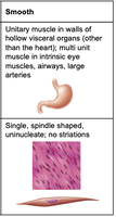
The human body contains three main types of muscle tissue, each with distinct structural and functional characteristics. The muscular system specifically refers to skeletal muscle, which is responsible for voluntary movements.
Skeletal Muscle: Attached to bones and skin, responsible for voluntary movements, striated appearance.
Cardiac Muscle: Found only in the heart, responsible for pumping blood, involuntary, striated.
Smooth Muscle: Located in walls of hollow organs, responsible for involuntary movements, non-striated.
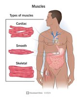
Skeletal Muscle
Location: Attached to bones or skin.
Cell Structure: Single, very long, cylindrical, multinucleate cells with obvious striations.
Function: Voluntary movement, rapid contraction, powerful but tires easily.
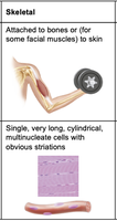
Cardiac Muscle
Location: Walls of the heart.
Cell Structure: Branching chains of cells; uni- or binucleate; striations.
Function: Involuntary contraction, pumps blood throughout the body.
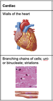
Smooth Muscle
Location: Walls of hollow visceral organs (e.g., stomach, airways, arteries).
Cell Structure: Single, spindle-shaped, uninucleate; no striations.
Function: Involuntary contraction, moves substances through internal passageways.
Functions of Muscles
Muscles perform several essential functions in the body:
Produce Movement: Responsible for locomotion and manipulation.
Maintain Posture: Stabilize body position.
Stabilize Joints: Support and reinforce joints.
Generate Heat: Muscle contraction produces heat, helping maintain body temperature.
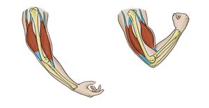
Skeletal Muscle Structure and Organization
Nerve and Blood Supply
Each skeletal muscle receives a nerve, artery, and vein. Contracting muscle fibers require large amounts of oxygen and nutrients, supplied by the blood vessels.
Connective Tissue Sheaths
Connective tissue sheaths support muscle cells and reinforce the whole muscle. There are three main layers:
Epimysium: Surrounds the entire muscle.
Perimysium: Surrounds bundles of muscle fibers (fascicles).
Endomysium: Surrounds individual muscle fibers.
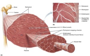
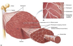
Muscle Attachments
Muscles span joints and attach to bones or other structures. Attachments can be direct (muscle attaches directly to bone) or indirect (muscle attaches via tendons or aponeuroses).
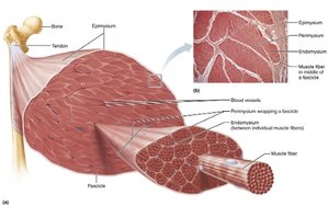
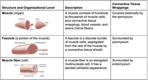
Organizational Levels of Skeletal Muscle
Skeletal muscle is organized into hierarchical levels:
Structure and Organizational Level | Description | Connective Tissue Wrappings |
|---|---|---|
Muscle (organ) | A muscle consists of hundreds to thousands of muscle cells, plus connective tissue wrappings, blood vessels, and nerves. | Covered externally by the epimysium |
Fascicle (portion of the muscle) | A fascicle is a discrete bundle of muscle cells, segregated from the rest of the muscle by a connective tissue sheath. | Surrounded by perimysium |
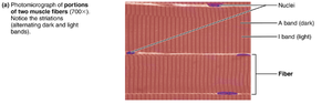
Muscle fiber (cell) | A muscle fiber is an elongated multinucleate cell; it has a banded (striated) appearance. | Surrounded by endomysium |
Skeletal Muscle Fibers
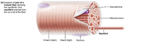
Skeletal muscle fibers are long, cylindrical cells containing multiple nuclei. The plasma membrane is called the sarcolemma, and the cytoplasm is called sarcoplasm. Sarcoplasm contains many glycosomes for glycogen storage and myoglobin for oxygen storage.
Specialized Structures
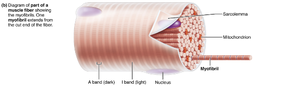
Myofibrils: Densely packed, rodlike elements responsible for muscle contraction.
Sarcoplasmic Reticulum: Network of smooth endoplasmic reticulum tubules surrounding each myofibril, regulates Ca2+ levels.
T Tubules: Tubes formed by protrusion of sarcolemma deep into cell interior, allow electrical signals to reach deep into muscle fiber.
Myofibrils: Structure and Function
Striations
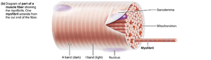
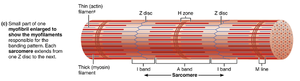
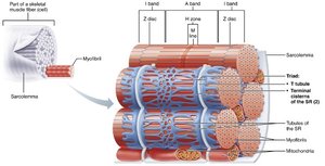
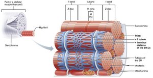
Striations are stripes formed from repeating series of dark (A bands) and light (I bands) along the length of each myofibril. The H zone is a lighter region in the middle of the A band, and the M line bisects the H zone vertically. The Z disc is a coin-shaped sheet of proteins on the midline of the I band.
Sarcomere
The sarcomere is the smallest contractile unit of a muscle fiber. It contains an A band with half of an I band at each end. Sarcomeres align end to end along myofibrils, like boxcars of a train.
Myofilaments
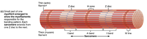
Myofilaments are the contractile proteins within the sarcomere. There are two main types:
Actin (thin filaments): Arranged in a hexagonal pattern around thick filaments.
Myosin (thick filaments): Surrounded by six thin filaments.
Molecular Composition of Myofilaments
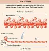
Thick Filaments: Composed of protein myosin, which contains two heavy and four light polypeptide chains. Heavy chains form the myosin tail, and light chains form the globular head. During contraction, heads link thick and thin filaments together, forming cross bridges.
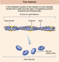
Thin Filaments: Composed of fibrous protein actin. G actin subunits bear active sites for myosin head attachment. Two F actin strands twist together to form a thin filament. Regulatory proteins tropomyosin and troponin are bound to actin.
Sarcoplasmic Reticulum and T Tubules
Sarcoplasmic Reticulum (SR)
The SR is a network of smooth endoplasmic reticulum tubules surrounding each myofibril. Most run longitudinally, and terminal cisterns form perpendicular cross channels at the A–I band junction. The SR regulates intracellular Ca2+ levels, storing and releasing Ca2+ as needed for muscle contraction.
T Tubules
T tubules are formed by protrusion of the sarcolemma deep into the cell interior. They greatly increase the muscle fiber’s surface area and allow electrical nerve transmissions to reach deep into the interior of each muscle fiber. The lumen of T tubules is continuous with the extracellular space.
Summary Table: Muscle Types
Muscle Type | Location | Cell Structure | Control |
|---|---|---|---|
Skeletal | Attached to bones/skin | Long, cylindrical, multinucleate, striated | Voluntary |
Cardiac | Heart | Branching, uni- or binucleate, striated | Involuntary |
Smooth | Walls of hollow organs | Spindle-shaped, uninucleate, non-striated | Involuntary |
Example: Skeletal muscles such as the biceps brachii allow voluntary movement of the forearm, while cardiac muscle contracts rhythmically to pump blood, and smooth muscle in the intestines moves food along the digestive tract.
Additional info: The notes have been expanded to include definitions, examples, and structural context for each muscle type and their organizational levels, as well as the molecular basis of muscle contraction.
