 Indietro
The Muscular System: Structure, Function, and Major Muscles
Guida di studio - Note intelligenti
 Appunti personalizzati basati sui tuoi materiali, ampliati con definizioni chiave, esempi e contesto.
The Muscular System
Introduction
The muscular system is essential for movement, posture, and many vital functions in the human body. This chapter focuses on skeletal muscles, their organization, actions, and the principles underlying their function.
Muscle Actions and Interactions
Functional Groups of Muscles
Prime Mover (Agonist): The main muscle responsible for a specific movement.
Antagonist: Muscle that opposes or reverses a movement; located on the opposite side of the joint from the agonist.
Synergist: Assists the prime mover by adding force or reducing unwanted movement.
Fixator: A type of synergist that stabilizes the origin of the prime mover.
Muscles can only pull, not push. What one muscle group does, another undoes.
Inferring Muscle Action by Position
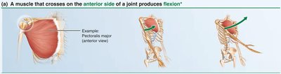
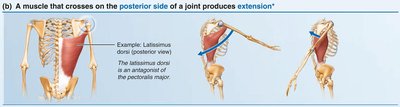
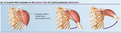
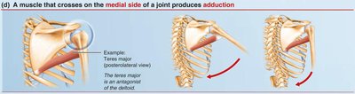
The action of a muscle can often be inferred by its position relative to the joint it crosses. For example, at the shoulder joint:
A muscle crossing on the anterior side produces flexion.
A muscle crossing on the posterior side produces extension.
A muscle crossing on the lateral side produces abduction.
A muscle crossing on the medial side produces adduction.
Note: These generalities do not apply to the knee and ankle due to limb rotation during development.
Naming Skeletal Muscles
Criteria for Naming Muscles
Location: Named for the bone or region (e.g., temporalis over the temporal bone).
Shape: Named for distinctive shapes (e.g., deltoid = triangle).
Size: Terms like maximus (largest), minimus (smallest), longus (long).
Direction of Fibers: Rectus (straight), transversus (right angles), oblique (at angles).
Number of Origins: Biceps (two origins), triceps (three origins).
Location of Attachments: Named for origin and insertion (e.g., sternocleidomastoid).
Action: Named for the movement produced (e.g., flexor, extensor).
Combination: Several criteria may be combined (e.g., extensor carpi radialis longus).
Fascicle Arrangements in Muscles
Patterns of Fascicle Arrangement
Fascicles are bundles of muscle fibers. Their arrangement affects muscle shape and function:
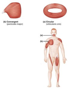
Circular: Fascicles arranged in concentric rings (e.g., orbicularis oris).
Convergent: Broad origin, fascicles converge toward a single tendon (e.g., pectoralis major).
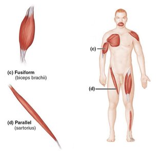
Parallel: Fascicles parallel to the long axis (e.g., sartorius).
Fusiform: Spindle-shaped with parallel fibers (e.g., biceps brachii).
Pennate: Short fascicles attach obliquely to a central tendon. Types include unipennate, bipennate, and multipennate.
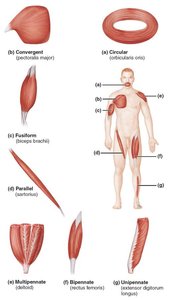
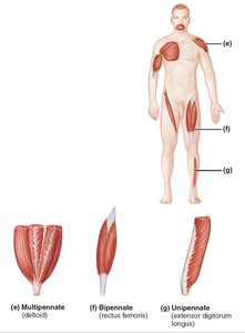
Functional Implications: Fascicle arrangement determines a muscle’s range of motion and power. Long, parallel fibers allow greater range but less power; pennate muscles are more powerful but have less range of motion.
Lever Systems in the Body
Basic Principles of Levers
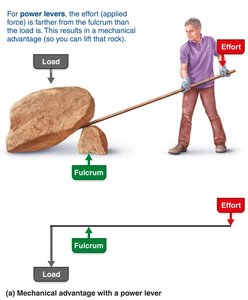
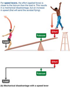
Most skeletal muscles use bones as levers to move loads. The three main components are:
Lever: Rigid bar (bone) that moves on a fixed point (fulcrum).
Fulcrum: The joint.
Effort: Force applied by muscle contraction.
Load: Resistance moved by the effort (bone, tissue, added weight).
Power Versus Speed Levers
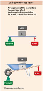
Mechanical Advantage (Power Lever): Load is close to fulcrum, effort is far. Small effort moves large load.
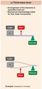
Mechanical Disadvantage (Speed Lever): Load is far from fulcrum, effort is close. Load moves rapidly over a large distance.
Classes of Levers
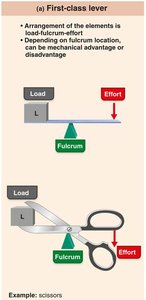
Class | Arrangement | Example | Advantage |
|---|---|---|---|
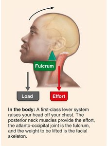
First-class | Fulcrum between load and effort | Seesaw, scissors, raising head off chest | Can be advantage or disadvantage |
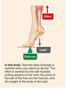
Second-class | Load between fulcrum and effort | Wheelbarrow, standing on tiptoe | Mechanical advantage (powerful) |
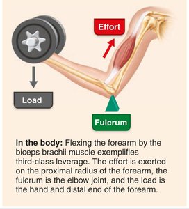
Third-class | Effort between fulcrum and load | Tweezers, most skeletal muscles (e.g., biceps brachii) | Mechanical disadvantage (speed and range) |
Major Skeletal Muscles of the Body
Overview of Major Muscles
There are over 600 skeletal muscles in the human body, grouped by function and location. Understanding their origin, insertion, action, and innervation is key to mastering anatomy.
Origin: Attachment point that moves least (usually proximal or medial).
Insertion: Attachment point that moves most (usually distal or lateral).
Action: The movement produced by muscle contraction.
Innervation: The nerve supplying the muscle.
Superficial Muscles: Anterior and Posterior Views
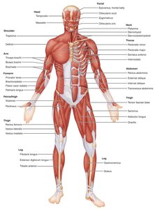
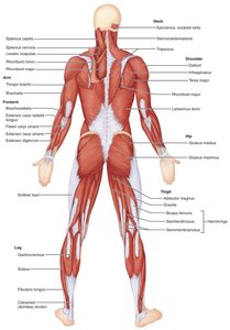
Muscles of the Head: Facial Expression
Facial muscles are unique because they insert into the skin, not bone, allowing for a wide range of expressions. All are innervated by the facial nerve (cranial nerve VII).
Epicranius (frontal and occipital bellies): Raises eyebrows, wrinkles forehead, pulls scalp backward.
Orbicularis oculi: Closes eye, blinking, squinting.
Zygomaticus: Raises corners of mouth (smiling).
Risorius: Pulls corner of lip laterally.
Orbicularis oris: Closes, purses lips (kissing, whistling).
Buccinator: Compresses cheek (whistling, sucking).
Platysma: Tenses skin of neck, depresses mandible.
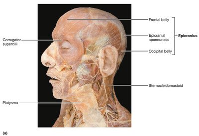
Example: The zygomaticus muscle is responsible for smiling by raising the lateral corners of the mouth.
*Additional info: For a complete understanding, students should also study muscle tables and diagrams for specific origins, insertions, and actions of each muscle group.*
