Indietro
The Muscular System: Structure, Types, and Function
Guida di studio - Note intelligenti
 Appunti personalizzati basati sui tuoi materiali, ampliati con definizioni chiave, esempi e contesto.
The Muscular System
Overview
The muscular system is essential for movement, posture, and various bodily functions. It is composed of three main types of muscle tissue: skeletal, cardiac, and smooth muscle. This section provides an in-depth look at the structure, function, and organization of muscles, focusing primarily on skeletal muscle as it relates to the human body's voluntary movements.
Types of Muscle
Classification of Muscle Tissue
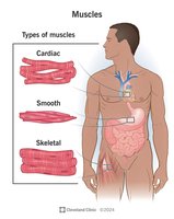
Skeletal Muscle: Attached to bones and skin, responsible for voluntary movements, and characterized by striations.
Cardiac Muscle: Found only in the heart, responsible for pumping blood, involuntary, and striated.
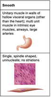
Smooth Muscle: Located in the walls of hollow organs (e.g., stomach, intestines), involuntary, and non-striated.
Key Structural and Functional Differences
Muscle Type | Location | Cell Shape & Nuclei | Striations | Control |
|---|---|---|---|---|
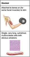
Skeletal | Attached to bones or skin | Long, cylindrical, multinucleate | Striated | Voluntary |
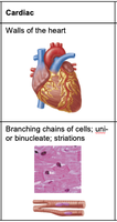
Cardiac | Walls of the heart | Branching, uni- or binucleate | Striated | Involuntary |
Smooth | Walls of hollow organs | Spindle-shaped, uninucleate | Non-striated | Involuntary |
Functions of Muscle
Major Roles of Muscle Tissue
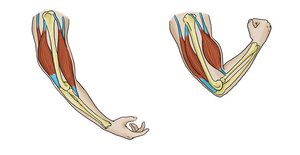
Produce Movement: Muscles are responsible for all voluntary and involuntary movements, including locomotion and manipulation of the environment.
Maintain Posture and Body Position: Continuous muscle contractions stabilize the body and maintain posture.
Stabilize Joints: Muscles reinforce and stabilize joints, preventing dislocation.
Generate Heat: Muscle contractions produce heat, which is important for maintaining body temperature.
Skeletal Muscle Structure and Organization
Nerve and Blood Supply
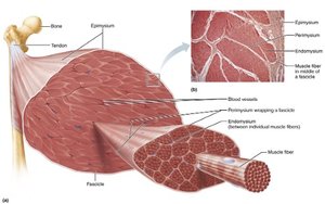
Each skeletal muscle receives a nerve, artery, and vein.
Muscle fibers require large amounts of oxygen and nutrients, delivered by blood vessels.
Connective Tissue Sheaths
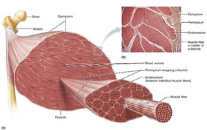
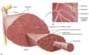
Each skeletal muscle and its fibers are covered by connective tissue sheaths that support and reinforce the muscle:
Epimysium: Surrounds the entire muscle.
Perimysium: Surrounds bundles of muscle fibers (fascicles).
Endomysium: Surrounds each individual muscle fiber.
Muscle Attachments
Muscles span joints and attach to bones or other structures via tendons (indirect) or directly to bone (direct).
Organizational Levels of Skeletal Muscle
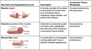
Skeletal muscle is organized into hierarchical levels:
Structure and Organizational Level | Description | Connective Tissue Wrappings |
|---|---|---|
Muscle (organ) | Hundreds to thousands of muscle cells, plus connective tissue, blood vessels, and nerves | Epimysium |
Fascicle (portion of muscle) | Discrete bundle of muscle cells, segregated by connective tissue sheath | Perimysium |
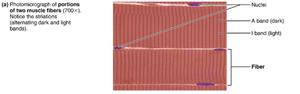
Muscle fiber (cell) | Elongated, multinucleate cell with striated appearance | Endomysium |
Skeletal Muscle Fibers
Structure of Muscle Fibers
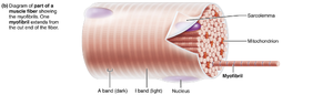
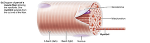
Long, cylindrical cells with multiple nuclei.
Sarcolemma: Plasma membrane of the muscle fiber.
Sarcoplasm: Cytoplasm of the muscle fiber, containing glycosomes (for glycogen storage) and myoglobin (for oxygen storage).
Specialized Structures
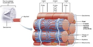
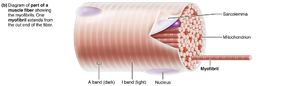
Myofibrils: Densely packed, rodlike elements responsible for muscle contraction.
Sarcoplasmic Reticulum (SR): Specialized endoplasmic reticulum that stores and releases calcium ions.
T Tubules: Invaginations of the sarcolemma that transmit electrical impulses deep into the muscle fiber.
Myofibrils and Striations
Striations and Sarcomeres
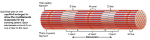
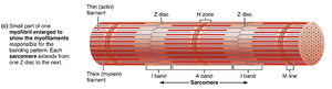
Striations: Alternating dark (A bands) and light (I bands) regions along the length of each myofibril.
Sarcomere: The smallest contractile unit of a muscle fiber, extending from one Z disc to the next.
Key regions: A band, H zone, M line, I band, Z disc.
Myofilaments: Actin and Myosin
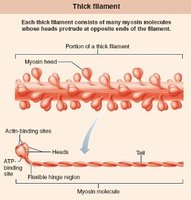
Thick filaments: Composed of myosin molecules, each with a tail and two heads that form cross-bridges during contraction.
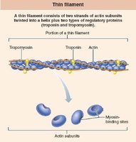
Thin filaments: Composed of actin, with regulatory proteins tropomyosin and troponin.
Arrangement: Each thick filament is surrounded by six thin filaments in a hexagonal pattern.
Sarcoplasmic Reticulum and T Tubules
Sarcoplasmic Reticulum (SR)
Network of smooth endoplasmic reticulum tubules surrounding each myofibril.
Terminal cisterns form cross channels at the A–I band junctions.
Functions in regulation of intracellular Ca2+ levels, storing and releasing calcium ions as needed for muscle contraction.
T Tubules
Formed by the sarcolemma protruding deep into the cell interior.
Increase the muscle fiber’s surface area and allow electrical nerve transmissions to reach deep into the muscle fiber.
Lumen is continuous with the extracellular space.