 Indietro
The Nervous System: Structure, Function, and Organization
Guida di studio - Note intelligenti
 Appunti personalizzati basati sui tuoi materiali, ampliati con definizioni chiave, esempi e contesto.
The Nervous System: Overview and Functions
General Functions of the Nervous System
The nervous system is responsible for monitoring internal and external environments, integrating sensory information, and coordinating voluntary and involuntary responses. It is essential for maintaining homeostasis and enabling complex behaviors.
Monitoring: Detects changes in the body and environment through sensory receptors.
Integration: Processes and interprets sensory input, deciding on appropriate responses.
Coordination: Initiates and regulates motor output to effectors (muscles and glands).
Divisions of the Nervous System
Central and Peripheral Nervous Systems
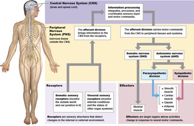
The nervous system is divided into the Central Nervous System (CNS) and Peripheral Nervous System (PNS), each with distinct roles:
CNS: Consists of the brain and spinal cord; responsible for information processing and integration.
PNS: Composed of all nervous tissue outside the CNS; connects the CNS to limbs and organs.
The PNS is further divided into:
Afferent Division: Carries sensory information to the CNS.
Efferent Division: Transmits motor commands from the CNS to effectors.
Somatic Nervous System (SNS): Controls voluntary movements of skeletal muscles.
Autonomic Nervous System (ANS): Regulates involuntary functions (e.g., heart rate, digestion).
Neural Tissue
Neurons and Neuroglia
Neural tissue consists of two main cell types: neurons and neuroglia (glial cells).
Neurons: The basic functional units responsible for communication via electrical and chemical signals.
Neuroglia: Support, protect, and nourish neurons; maintain the environment around neurons.
Structure of a Neuron
Neurons have specialized structures for receiving, processing, and transmitting information:
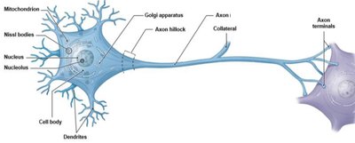
Dendrites: Receive incoming signals.
Cell Body (Soma): Contains the nucleus and organelles.
Axon Hillock: Site where action potentials are initiated.
Axon: Conducts action potentials away from the cell body.
Axon Terminals: Release neurotransmitters to communicate with other cells.
Structural and Functional Classification of Neurons
Multipolar Neurons: Multiple dendrites, one axon; most common in CNS; all motor neurons controlling skeletal muscles.
Unipolar Neurons: Dendrites and axon are continuous; most sensory neurons in PNS.
Bipolar Neurons: One dendrite, one axon; rare, found in special sense organs.
Sensory (Afferent) Neurons: Transmit sensory information to the CNS.
Motor (Efferent) Neurons: Carry commands from the CNS to effectors.
Interneurons: Connect neurons within the CNS; involved in processing and integration.
Neuroglia (Glial Cells)
Neuroglia in the CNS
Astrocytes: Maintain blood-brain barrier, provide structural support, repair tissue.
Oligodendrocytes: Form myelin sheaths around CNS axons, increasing conduction speed.
Microglia: Phagocytic cells that remove debris and pathogens.
Ependymal Cells: Line ventricles, produce and circulate cerebrospinal fluid (CSF).
Neuroglia in the PNS
Satellite Cells: Support neuron cell bodies in ganglia.
Schwann Cells: Myelinate axons in the PNS; essential for nerve regeneration.
Organization of Neurons in the Nervous System
Gray and White Matter
Gray Matter: Contains neuron cell bodies, dendrites, and unmyelinated axons.
White Matter: Composed of myelinated axons; forms tracts and columns in the CNS.
In the PNS, neuron cell bodies are grouped in ganglia, and axons are bundled into nerves.
Membrane Potential and Action Potentials
Resting Membrane Potential
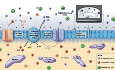
Neurons maintain a resting membrane potential, typically around -70 mV, due to differences in ion concentrations across the plasma membrane and selective permeability.
High Na+ and Cl- outside the cell
High K+ and negatively charged proteins (Pr-) inside the cell
Sodium-potassium exchange pump: Maintains ion gradients by pumping 3 Na+ out and 2 K+ in per ATP hydrolyzed.
Changes in Membrane Potential
Stimuli that alter membrane permeability to Na+ or K+ can change the membrane potential, leading to graded or action potentials.
Graded Potentials: Local changes in membrane potential.
Action Potentials: Rapid, large changes in membrane potential that propagate along axons.
Action Potential Phases
Depolarization to Threshold: Membrane potential becomes less negative.
Activation of Sodium Channels: Rapid influx of Na+ causes further depolarization.
Inactivation of Sodium Channels, Activation of Potassium Channels: K+ exits the cell, repolarizing the membrane.
Return to Resting Potential: K+ channels close, membrane returns to resting state.
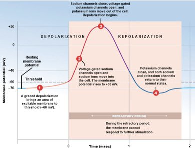
Refractory Period: Time during which the neuron cannot fire another action potential.
Synapses and Neurotransmitters
Neuroeffector Junctions and Synaptic Transmission
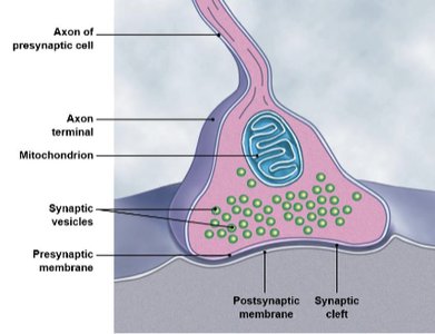
Synapses are specialized junctions where neurons communicate with other neurons or effectors via neurotransmitters.
Presynaptic Neuron: Releases neurotransmitter into the synaptic cleft.
Postsynaptic Neuron: Receives the signal via receptors.
Types of Neurotransmitters
Acetylcholine (ACh): Common in CNS and PNS; usually excitatory.
Norepinephrine: Found in brain and ANS; usually excitatory.
Dopamine, GABA, Serotonin: Found in CNS; often inhibitory.
The Central Nervous System (CNS)
Meninges
The CNS is protected by three connective tissue layers called meninges:
Dura Mater: Outermost, tough layer; forms dural folds and sinuses.
Arachnoid Mater: Middle layer; contains subarachnoid space filled with CSF.
Pia Mater: Innermost, delicate layer; adheres to CNS surface, highly vascularized.
Spinal Cord Anatomy
31 segments: Each gives rise to a pair of spinal nerves (8 cervical, 12 thoracic, 5 lumbar, 5 sacral, 1 coccygeal).
Central Canal: Contains CSF.
Gray Matter: Contains neuron cell bodies; organized into horns (anterior, lateral, posterior).
White Matter: Contains myelinated axons; organized into columns and tracts.
The Brain
Major Regions of the Brain
Cerebrum: Conscious thought, memory, sensory processing, voluntary movement.
Diencephalon: Sensory relay, autonomic functions, hormone production.
Midbrain: Visual and auditory processing, consciousness regulation.
Pons: Motor control, links cerebellum with other brain regions.
Medulla Oblongata: Autonomic centers for heart rate, respiration.
Cerebellum: Coordination of movement, balance, and posture.
Cerebral Cortex and Lobes
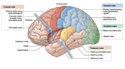
The cerebral cortex is divided into lobes, each with specialized functions:
Frontal Lobe: Voluntary movement, planning, reasoning.
Parietal Lobe: Sensory processing (touch, pressure, pain).
Occipital Lobe: Visual processing.
Temporal Lobe: Auditory and olfactory processing.
Insula: Taste and visceral sensation.
Hemispheric Specialization
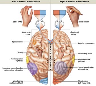
The left and right cerebral hemispheres have specialized functions and control opposite sides of the body.
Left Hemisphere: Language, logic, analytical tasks.
Right Hemisphere: Spatial abilities, creativity, recognition of faces and patterns.
Basal Nuclei
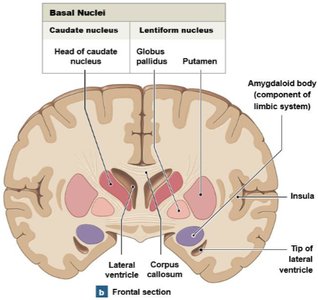
Basal nuclei are deep gray matter structures involved in the coordination of movement and processing of sensory information outside conscious awareness.
The Diencephalon
Components and Functions
Epithalamus: Contains pineal gland; secretes melatonin for circadian rhythm regulation.
Thalamus: Relay station for sensory information; filters and directs signals to the cortex.
Hypothalamus: Regulates autonomic functions, hormone secretion, hunger, thirst, temperature, and circadian rhythms.
Brainstem and Cerebellum
Midbrain, Pons, and Medulla Oblongata
Midbrain: Integrates sensory information, regulates motor output, contains reflex centers.
Pons: Controls respiration, links cerebellum with other brain regions.
Medulla Oblongata: Contains cardiovascular and respiratory centers; connects brain and spinal cord.
Cerebellum
Coordinates voluntary and involuntary movements, balance, and posture.
Stores learned motor patterns.
Peripheral Nervous System (PNS)
Cranial and Spinal Nerves
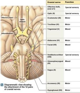
Cranial Nerves: 12 pairs; originate from the brain; sensory, motor, or mixed functions.
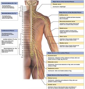
Spinal Nerves: 31 pairs; originate from the spinal cord; grouped by vertebral region.
Dermatomes
Each spinal nerve innervates a specific region of skin called a dermatome, useful in clinical diagnosis of nerve damage.
Reflexes
Reflex Arc
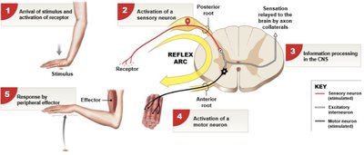
A reflex is a rapid, automatic response to a stimulus, involving a specific neural pathway called a reflex arc.
Stimulus → Sensory neuron → Interneuron (CNS) → Motor neuron → Effector
Autonomic Nervous System (ANS)
General Organization
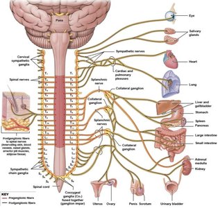
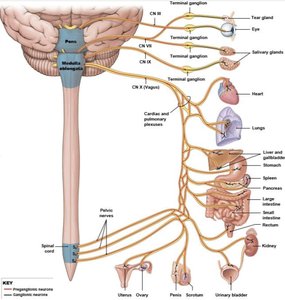
The ANS regulates involuntary functions and is divided into sympathetic and parasympathetic divisions.
Preganglionic Neurons: Originate in CNS, synapse in autonomic ganglia.
Postganglionic Neurons: Originate in ganglia, innervate target organs.
Sympathetic Division (Fight or Flight)
Prepares the body for stress or emergency situations.
Increases heart rate, dilates pupils, inhibits digestion, stimulates sweat glands.
Neurotransmitter: Norepinephrine (adrenergic).
Parasympathetic Division (Rest and Digest)
Promotes relaxation, digestion, and energy storage.
Decreases heart rate, constricts pupils, stimulates digestive activity.
Neurotransmitter: Acetylcholine (cholinergic).
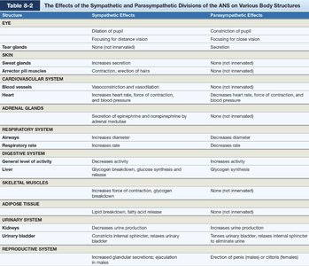
Comparison of Sympathetic and Parasympathetic Effects
Structure | Sympathetic Effects | Parasympathetic Effects |
|---|---|---|
Eye | Dilation of pupil | Constriction of pupil |
Heart | Increases rate and force | Decreases rate and force |
Digestive System | Decreases activity | Increases activity |
Respiratory System | Dilates airways | Constricts airways |
Adrenal Glands | Secretion of epinephrine/norepinephrine | None |
Urinary System | Decreases urine production | Increases urine production |
Reproductive System | Increases glandular secretion, ejaculation | Erection of penis/clitoris |
Skin (sweat glands) | Increases secretion | None |
Additional info: The ANS is crucial for maintaining homeostasis by balancing the opposing actions of the sympathetic and parasympathetic divisions.
