 Indietro
The Nervous System: Structure, Function, and Cellular Organization
Guida di studio - Note intelligenti
 Appunti personalizzati basati sui tuoi materiali, ampliati con definizioni chiave, esempi e contesto.
Nervous System Overview
General Functions and Importance
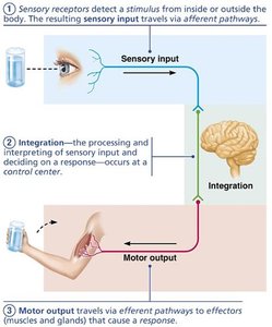
The nervous system is the master controlling and communicating system of the body. It receives, integrates, and responds to information through rapid and specific electrical and chemical signals, usually resulting in immediate responses.
Sensory input: Detection of internal and external stimuli.
Integration: Processing and interpretation of sensory input, leading to decision-making.
Motor output: Activation of effector organs (muscles and glands) to produce a response.
Anatomical Subdivisions of the Nervous System
Central and Peripheral Nervous Systems
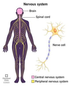
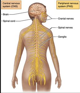
The nervous system is divided into two principal parts:
Central Nervous System (CNS): Consists of the brain and spinal cord; serves as the integrative and control center.
Peripheral Nervous System (PNS): Composed mainly of nerves extending from the brain and spinal cord, including cranial and spinal nerves, and ganglia. The PNS connects the CNS to the rest of the body.
Functional Divisions of the PNS
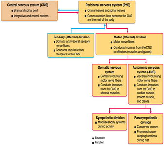
The PNS is functionally divided into:
Sensory (afferent) division: Conducts impulses from sensory receptors to the CNS.
Motor (efferent) division: Conducts impulses from the CNS to effectors (muscles and glands).
Motor Division Subdivisions
The motor division of the PNS is further subdivided into:
Somatic Nervous System: Controls voluntary movements by conducting impulses to skeletal muscles.
Autonomic Nervous System (ANS): Controls involuntary functions by conducting impulses to cardiac muscle, smooth muscle, and glands. The ANS is subdivided into:
Sympathetic division: Mobilizes body systems during activity (fight or flight).
Parasympathetic division: Conserves energy and promotes housekeeping functions during rest.
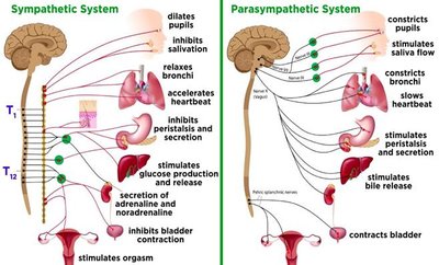
Nervous Tissue: Cellular Components
Principal Cell Types
Nervous tissue consists of two main cell types:
Neuroglia (glial cells): Support, protect, and nourish neurons; play a central role in nervous system health.
Neurons (nerve cells): Excitable cells that transmit electrical signals.
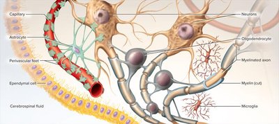
Neuroglial Cells of the CNS
There are four types of glial cells in the CNS:
Astrocytes: Most abundant; support neurons, regulate the blood-brain barrier, and maintain extracellular environment.
Microglia: Small, phagocytic cells; act as immune cells, removing pathogens and debris.
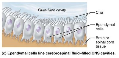
Ependymal cells: Line fluid-filled cavities; produce and circulate cerebrospinal fluid (CSF).
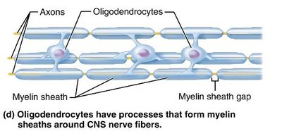
Oligodendrocytes: Form myelin sheaths around CNS axons, increasing signal transmission speed.
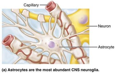
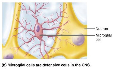
Neuroglial Cells of the PNS
The PNS contains two types of glial cells:
Satellite cells: Surround neuron cell bodies in ganglia; similar to astrocytes in function.
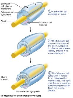
Schwann cells: Form myelin sheaths around PNS axons; vital for regeneration of damaged nerve fibers.
Neurons: Structure and Function
Basic Structure of a Neuron
Neurons are large, highly specialized cells with unique properties:
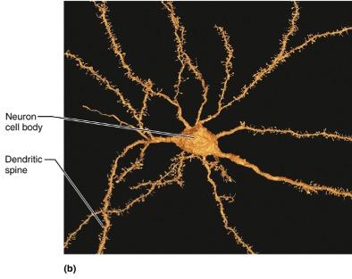
Cell body (soma): Contains the nucleus and metabolic machinery; receptive region for signals.
Dendrites: Branching arms that receive signals from other neurons; dendritic spines increase connectivity and are associated with learning and memory.
Axon: Single, long process that transmits impulses away from the cell body; may branch and end in axon terminals.
Myelin Sheath
The myelin sheath is a white, fatty substance that coats many axons, providing:
Protection and electrical insulation
Increased speed of nerve impulse transmission
Myelinated axons: Segmented sheath surrounds most long or large-diameter axons.
Non-myelinated axons: Lack sheath; conduct impulses more slowly.
Myelin Sheath in the PNS
Formed by Schwann cells wrapping around the axon in a jelly roll fashion.
Each Schwann cell forms one segment of myelin sheath.
Myelin sheath gaps (nodes of Ranvier) are sites where axon collaterals can emerge.
Myelin Sheath in the CNS
Formed by processes of oligodendrocytes, not whole cells.
Each oligodendrocyte can wrap up to 60 axons at once.
Myelin sheath gaps are present; no outer collar of perinuclear cytoplasm.
Thinnest fibers are unmyelinated but covered by long extensions of adjacent neuroglia.
Classification of Neurons
Structural Classification
Neurons are classified based on the number of processes arising from the cell body:
Multipolar: One axon, multiple dendrites; most common type.
Bipolar: One axon, one dendrite; found in sense organs.
Unipolar: Single process branches into a T; dendrites receive signals from skin and joints, axon leads to spinal cord.
Functional Classification
Neurons are grouped by the direction of nerve impulse travel relative to the CNS:
Sensory (afferent) neurons: Transmit impulses from sensory receptors toward the CNS; almost all are unipolar, cell bodies located in PNS ganglia.
Motor (efferent) neurons: Carry outgoing signals from the CNS to effectors; usually multipolar.
Interneurons (association neurons): Perform integrative functions; only found in the CNS; most abundant and usually multipolar.
Summary Table: Structural and Functional Classes of Neurons
Structural Class | Processes | Location | Functional Class |
|---|---|---|---|
Multipolar | 1 axon, multiple dendrites | CNS, most neurons | Motor, interneurons |
Bipolar | 1 axon, 1 dendrite | Sensory organs | Sensory |
Unipolar | Single process (T-shaped) | PNS, ganglia | Sensory |
Key Terms and Concepts
Neuron: Excitable cell transmitting electrical signals.
Neuroglia: Supportive cells in nervous tissue.
Myelin sheath: Insulating layer around axons.
Central Nervous System (CNS): Brain and spinal cord.
Peripheral Nervous System (PNS): Nerves and ganglia outside CNS.
Somatic Nervous System: Voluntary control of skeletal muscles.
Autonomic Nervous System (ANS): Involuntary control of organs.
Sympathetic Division: Mobilizes body for activity.
Parasympathetic Division: Conserves energy, promotes rest.
Equations and Academic Context
Resting Membrane Potential (for advanced context)
The resting membrane potential of a neuron is determined by the distribution of ions across the membrane and can be calculated using the Nernst equation: Additional info: This equation is provided for advanced students interested in the biophysical basis of neuronal signaling.
