 Indietro
Chapter 16- Lecture 3
Guida di studio - Note intelligenti
 Appunti personalizzati basati sui tuoi materiali, ampliati con definizioni chiave, esempi e contesto.
The Autonomic Nervous System
Overview of the Autonomic Nervous System (ANS)
The Autonomic Nervous System (ANS) is a division of the peripheral nervous system responsible for involuntary physiological functions. It regulates vital processes such as heart rate, digestion, respiratory rate, pupillary response, and more. The ANS is divided into the sympathetic and parasympathetic divisions, which often have opposing effects on target organs.
Sympathetic Division: Prepares the body for 'fight or flight' responses.
Parasympathetic Division: Promotes 'rest and digest' activities, conserving energy and maintaining resting metabolic rate.
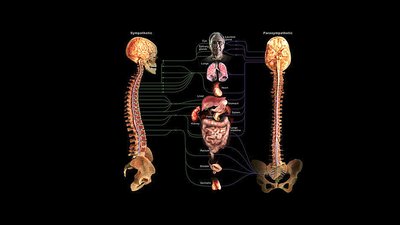
Parasympathetic Division (Craniosacral Division)
Structure and Organization
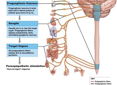
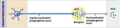
The parasympathetic division is also known as the craniosacral division due to its origin in the brainstem and sacral segments of the spinal cord. It is characterized by long preganglionic fibers and short postganglionic fibers, with ganglia located near or within target organs.
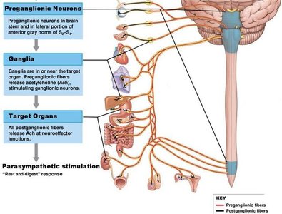
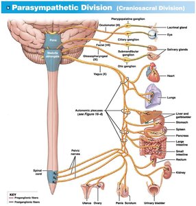
Preganglionic Neurons: Originate in the brainstem (cranial nerves III, VII, IX, X) and sacral spinal cord (S2-S4).
Ganglia: Terminal ganglia (near target organs) and intramural ganglia (within target organs).
Postganglionic Neurons: Short fibers that innervate target tissues.
Parasympathetic Ganglia
Parasympathetic ganglia are classified as terminal or intramural ganglia. Terminal ganglia are associated with cranial nerves and control visceral structures in the head, while intramural ganglia are embedded within the walls of target organs.
Terminal Ganglia: Associated with cranial nerves III (oculomotor), VII (facial), IX (glossopharyngeal).
Intramural Ganglia: Located within organs such as the heart, lungs, digestive tract, and urinary bladder.

The Vagus Nerve (Cranial Nerve X)
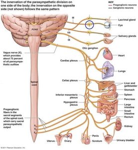
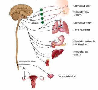
The vagus nerve is the most significant parasympathetic nerve, providing approximately 80% of all parasympathetic outflow. It innervates structures in the neck, thoracic cavity, and abdominopelvic cavities, influencing heart rate, digestion, and other vital functions.
Functions: Slows heart rate, stimulates digestive activity, regulates respiratory rate, and more.
Clinical Relevance: Dysfunction can affect multiple organ systems.


Functions of the Parasympathetic Division
Rest and Digest Response
The parasympathetic division conserves energy and maintains resting metabolic rate. It is active during periods of rest, promoting digestion, nutrient absorption, and waste elimination.
Pupil constriction: Facilitates focusing on nearby objects.
Stimulates tear and saliva production: Essential for eye and oral health.
Slows heart rate: Maintains cardiovascular homeostasis.
Stimulates peristalsis and secretion: Enhances digestive function.
Contracts bladder: Promotes urination.

Mechanisms of Parasympathetic Neurotransmitter Release
Acetylcholine (ACh) Release and Receptors
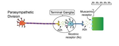
All parasympathetic neurons, both preganglionic and postganglionic, release acetylcholine (ACh). The effects on target organs depend on the type of receptor present and the nature of the second messenger system activated.
Nicotinic Receptors: Located on postganglionic neurons; always excitatory.
Muscarinic Receptors: Located on target organs; effects can be excitatory or inhibitory depending on receptor subtype and enzyme activation.
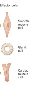
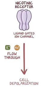
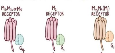
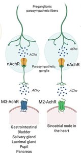
Muscarinic Receptor Subtypes and Effects
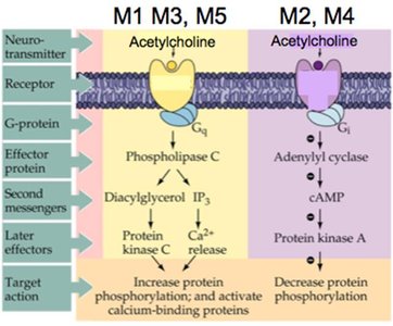
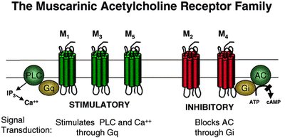
Muscarinic receptors are G-protein coupled receptors with several subtypes (M1, M2, M3, M4, M5). Their effects vary:
M1, M3, M5: Excitatory, stimulate phospholipase C, increase intracellular Ca2+.
M2, M4: Inhibitory, decrease cAMP, reduce protein phosphorylation.
Parasympathetic Agonists and Antagonists
Pharmacological Modulation
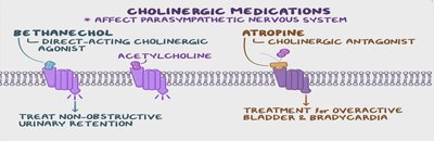
Drugs can mimic (agonists) or block (antagonists) parasympathetic activity. For example, methacholine acts like acetylcholine and is used to treat tachycardia, but may cause increased salivation, difficulty breathing, and other side effects.
Agonists: Stimulate parasympathetic responses (e.g., increased salivation, bronchoconstriction).
Antagonists: Inhibit parasympathetic responses (e.g., atropine for bradycardia or overactive bladder).
Comparison of Sympathetic and Parasympathetic Divisions
Key Differences
The sympathetic and parasympathetic divisions differ in their organization, neurotransmitters, receptor types, and effects on target organs.
Feature | Sympathetic | Parasympathetic |
|---|---|---|
Origin | Thoracolumbar (T1-L2) | Craniosacral (brainstem, S2-S4) |
Preganglionic Fiber Length | Short | Long |
Postganglionic Fiber Length | Long | Short |
Neurotransmitter (Postganglionic) | Norepinephrine | Acetylcholine |
Receptor Types | Adrenergic | Muscarinic |
Main Effects | Fight or flight | Rest and digest |
Summary
The parasympathetic division of the ANS is essential for maintaining homeostasis during rest, promoting digestion, and regulating organ function. Its effects are mediated by acetylcholine acting on muscarinic and nicotinic receptors, with clinical relevance in pharmacology and disease states.
