 Indietro
The Respiratory System: Functional Anatomy and Physiology
Guida di studio - Note intelligenti
 Appunti personalizzati basati sui tuoi materiali, ampliati con definizioni chiave, esempi e contesto.
The Respiratory System
Overview and Chapter Structure
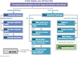
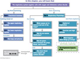
The respiratory system supplies cells with oxygen and eliminates carbon dioxide. It is divided into two main parts: Functional Anatomy and Respiratory Physiology. Functional anatomy covers the structures involved in respiration, while physiology explores the mechanisms and control of breathing.
Functional Anatomy of the Respiratory System
Four Processes of Respiration
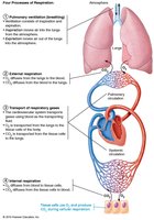
Respiration involves four essential processes:
Pulmonary Ventilation: Movement of air into and out of the lungs.
External Respiration: Gas exchange between lungs and blood.
Transport of Respiratory Gases: Movement of gases via blood between lungs and tissues.
Internal Respiration: Gas exchange between blood and tissue cells.
Major Structures of the Respiratory System
The respiratory system is divided into the upper and lower respiratory tracts, each with distinct anatomical features and functions.
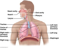
Upper Respiratory System
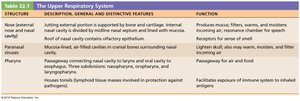
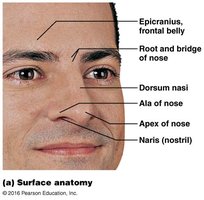
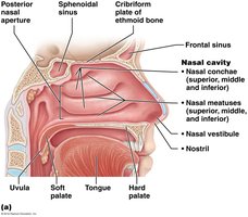
Nose/Nasal Cavity: Airway for respiration, moistens and warms air, filters particles, and houses olfactory receptors.
Paranasal Sinuses: Lighten skull, moisten air, and aid in resonance.
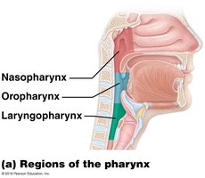
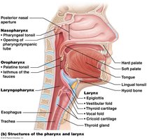
Pharynx: Passageway for air and food; includes nasopharynx, oropharynx, and laryngopharynx.
Lower Respiratory System
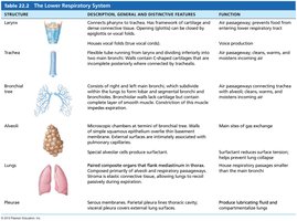
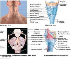
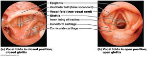
Larynx: Connects pharynx to trachea, houses vocal cords, and produces sound.
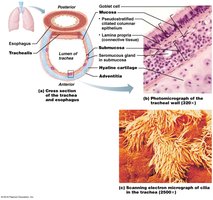
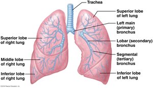
Trachea: Windpipe; passageway for air to the bronchi.
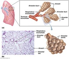
Bronchial Tree: Branching system of bronchi and bronchioles leading to alveoli.
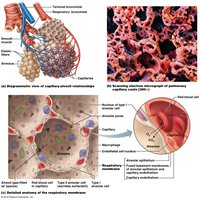
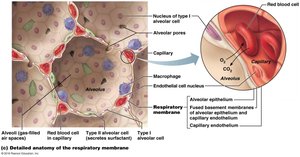
Alveoli: Site of gas exchange; contains Type I and Type II cells.
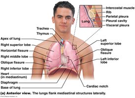
Lungs: Main organs of respiration, divided into lobes and segments.
Pleura: Double-layered membrane surrounding lungs, reduces friction.
Gross Anatomy of the Lungs and Pleurae
Lobes: Right lung has three lobes; left lung has two.
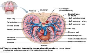
Pleurae: Parietal and visceral pleurae surround the lungs and create a pleural cavity.
Root, Hilum, and Cardiac Notch: Key anatomical landmarks for blood vessel and bronchial entry.
Respiratory Physiology
Pulmonary Ventilation
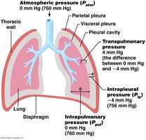
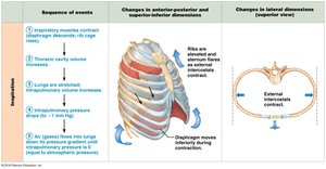
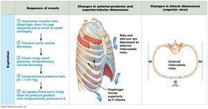
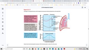
Pulmonary ventilation is the process of moving air into and out of the lungs. It is governed by pressure changes within the thoracic cavity.
Inspiration: Air flows into the lungs as thoracic volume increases and pressure decreases.
Expiration: Air leaves the lungs as thoracic volume decreases and pressure increases.
Atmospheric Pressure: At sea level, 760 mm Hg (1 atm).
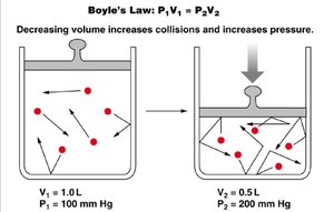
Boyle’s Law: At constant temperature, pressure and volume are inversely related:
Airway Resistance and Surfactant
Airway Resistance: Resistance is highest in medium-sized bronchi and decreases in smaller bronchioles.
Surfactant: Produced by Type II alveolar cells, reduces surface tension and prevents alveolar collapse.
Lung Compliance
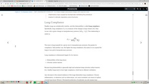
Lung compliance refers to the ability of the lungs to stretch and expand. It is calculated as:
Formula:
High compliance means easier expansion; low compliance indicates stiffness.
Factors: Distensibility of lung tissue and alveolar surface tension (overcome by surfactant).
Respiratory Volumes and Capacities
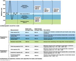
Respiratory volumes are measured using a spirometer. Key volumes include:
Tidal Volume (TV): Amount of air inhaled or exhaled per breath.
Inspiratory Reserve Volume (IRV): Extra air inhaled after a normal inspiration.
Expiratory Reserve Volume (ERV): Extra air exhaled after a normal expiration.
Residual Volume (RV): Air remaining in lungs after maximal exhalation.
Dead Space and Alveolar Ventilation
Anatomical Dead Space: Air in conducting passageways not involved in gas exchange (~150 mL).
Total Dead Space: Sum of all nonuseful air volume.
Alveolar Ventilation Rate (AVR):
Gas Exchange: External and Internal Respiration
External Respiration: Gas exchange between alveoli and blood.
Internal Respiration: Gas exchange between blood and tissues.
Dalton’s Law: Total pressure of a gas mixture is the sum of partial pressures.
Henry’s Law: Gas dissolves in liquid in proportion to its partial pressure.
Transport of Gases
Oxygen: Mostly transported by hemoglobin in RBCs; forms oxyhemoglobin.
Carbon Dioxide: Transported dissolved in plasma, bound to hemoglobin, or as bicarbonate ion.
Control of Respiration
Neural Controls: Respiratory centers in the brainstem regulate breathing rhythm.
Chemical Factors: CO2, O2, and pH levels influence respiratory rate and depth.
Exercise, Altitude, and Homeostatic Imbalances
Exercise: Increases ventilation due to neural and psychological stimuli.
Altitude: Lower partial pressures increase ventilation.
Homeostatic Imbalances: Includes COPD, asthma, tuberculosis, lung cancer, sinusitis, laryngitis, pleurisy, atelectasis, pneumothorax, oxygen toxicity, and hypoxia.
Summary Table: Upper and Lower Respiratory System Structures
Structure | Description | Function |
|---|---|---|
Nose/Nasal Cavity | Airway, moistens, warms, filters air | Respiration, olfaction, speech |
Paranasal Sinuses | Air-filled cavities | Lighten skull, moisten air |
Pharynx | Muscular tube | Passage for air and food |
Larynx | Connects pharynx to trachea | Voice production |
Trachea | Windpipe | Air passage |
Bronchial Tree | Branching bronchi | Air passage |
Alveoli | Site of gas exchange | Gas exchange |
Lungs | Main organ | Respiration |
Pleura | Membrane | Reduces friction |
Key Equations
Boyle’s Law:
Gas Flow:
Lung Compliance:
Alveolar Ventilation Rate:
Conclusion
The respiratory system is essential for gas exchange, maintaining homeostasis, and supporting cellular metabolism. Understanding its anatomy and physiology is crucial for recognizing normal function and disease states.
