 Indietro
The Respiratory System: Structure, Function, and Physiology
Guida di studio - Note intelligenti
 Appunti personalizzati basati sui tuoi materiali, ampliati con definizioni chiave, esempi e contesto.
Respiratory System Overview
Functions of the Respiratory System
The respiratory system is essential for gas exchange, supplying oxygen to the body and removing carbon dioxide. It also plays roles in sound production, olfaction (smell), and protection of the airways.
Oxygen Intake & Carbon Dioxide Removal: Delivers O2 to cells and removes CO2 waste.
Sound Production: Air movement over vocal folds enables speech.
Olfaction: Air passes over olfactory receptors in the nasal cavity.
Protection: Coughing and sneezing clear airways of debris.
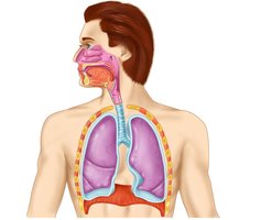
Mechanics of Breathing (Ventilation)
Inhalation and Exhalation
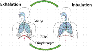
Breathing consists of two phases: inhalation (inspiration) and exhalation (expiration). These processes are driven by changes in thoracic cavity volume and pressure, primarily through the action of the diaphragm and intercostal muscles.
Inhalation: Diaphragm contracts and moves downward, rib cage expands, thoracic volume increases, and air flows into the lungs.
Exhalation: Diaphragm relaxes and moves upward, rib cage contracts, thoracic volume decreases, and air is expelled from the lungs.
Gas Exchange
External and Internal Respiration
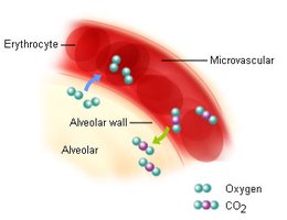
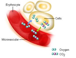
Gas exchange occurs in two main locations: the lungs (external respiration) and the tissues (internal respiration).
External Respiration: Exchange of O2 and CO2 between alveoli and pulmonary capillaries.
Internal Respiration: Exchange of O2 and CO2 between systemic capillaries and body tissues.
Anatomy of the Respiratory System
Conducting Zone vs. Respiratory Zone
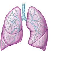
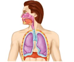
The respiratory system is divided into the conducting zone (air passageways) and the respiratory zone (site of gas exchange).
Conducting Zone: Includes nasal cavity, pharynx, larynx, trachea, bronchi, and bronchioles. Functions to transport, warm, humidify, and filter air.
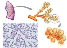
Respiratory Zone: Includes respiratory bronchioles, alveolar ducts, and alveoli. Site of external gas exchange.
Nasal Cavity and Paranasal Sinuses
The nasal cavity filters, warms, and humidifies incoming air. Paranasal sinuses lighten the skull and further condition the air.
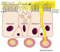
Nasal Cavity: Lined with pseudostratified ciliated columnar epithelium and goblet cells that secrete mucus.
Paranasal Sinuses: Air-filled spaces in the frontal, ethmoid, sphenoid, and maxillary bones.

Histology of the Respiratory Tract
Respiratory Epithelium
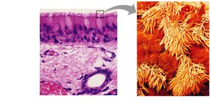
The respiratory tract is lined by specialized epithelium that varies by region:
Pseudostratified Ciliated Columnar Epithelium: Found in the nasal cavity, trachea, and bronchi; contains goblet cells for mucus production.
Stratified Squamous Epithelium: Found in areas subject to abrasion, such as the oropharynx and laryngopharynx.
Pharynx and Larynx
Pharynx
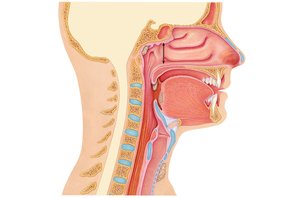
The pharynx is a muscular tube that connects the nasal cavity to the larynx and esophagus. It is divided into three regions:
Nasopharynx: Air passageway lined with pseudostratified ciliated columnar epithelium.
Oropharynx: Passageway for food and air, lined with stratified squamous epithelium.
Laryngopharynx: Passageway for food and air, lined with stratified squamous epithelium.
Larynx
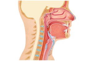
The larynx is responsible for air passage and voice production. It contains the vocal folds (true vocal cords) and is supported by several cartilages, including the thyroid, cricoid, and arytenoid cartilages.
Epiglottis: Elastic cartilage that covers the laryngeal inlet during swallowing.
Vocal Folds: Vibrate to produce sound; length and thickness determine pitch.
Lower Respiratory Tract
Trachea and Bronchial Tree
The trachea is a flexible tube supported by C-shaped hyaline cartilage rings. It branches into the primary bronchi, which further divide into secondary and tertiary bronchi, and finally into bronchioles.
Trachea: Lined with pseudostratified ciliated columnar epithelium and goblet cells.
Bronchi: Cartilage plates replace rings as bronchi branch; epithelium transitions from pseudostratified to simple columnar and then to simple cuboidal in bronchioles.
Bronchioles: Lack cartilage; smooth muscle regulates airflow.
Respiratory Zone and Alveoli
Alveolar Structure and Gas Exchange
The respiratory zone consists of respiratory bronchioles, alveolar ducts, and alveoli. Alveoli are the primary sites of gas exchange, surrounded by capillaries and composed of two main cell types.
Type I Alveolar Cells: Simple squamous cells forming the respiratory membrane.
Type II Alveolar Cells: Cuboidal cells that secrete surfactant to reduce surface tension.
Alveolar Macrophages: Remove debris and pathogens.
Respiratory Membrane
The respiratory membrane is the thin barrier through which gas exchange occurs between alveolar air and blood in capillaries. It consists of:
Alveolar epithelium (simple squamous)
Capillary endothelium (simple squamous)
Fused basal laminae between the two cell layers
Pleurae and Lung Structure
Pleural Membranes
The lungs are enclosed by serous membranes called pleurae:
Visceral Pleura: Covers the surface of the lungs.
Parietal Pleura: Lines the thoracic cavity.
Pleural Cavity: Space between the pleurae, filled with serous fluid to reduce friction during breathing.
Clinical Correlations
Respiratory Distress Syndrome
Premature infants may lack sufficient surfactant, making it difficult to inflate alveoli and leading to respiratory distress.
Emphysema and COPD
Chronic exposure to irritants (e.g., cigarette smoke) can cause inflammation, excess mucus, and destruction of alveolar walls, resulting in emphysema and chronic obstructive pulmonary disease (COPD).
Summary Table: Conducting vs. Respiratory Zone
Zone | Main Structures | Function | Epithelium |
|---|---|---|---|
Conducting | Nasal cavity, pharynx, larynx, trachea, bronchi, bronchioles | Transport, warm, humidify, filter air | Pseudostratified ciliated columnar (upper), simple columnar/cuboidal (lower) |
Respiratory | Respiratory bronchioles, alveolar ducts, alveoli | Gas exchange | Simple squamous (Type I), cuboidal (Type II) |
Key Equations
Gas Exchange (Fick's Law):
Boyle's Law (Breathing Mechanics):
Additional info: This law explains how changes in thoracic volume lead to changes in pressure, driving air movement during breathing.
