 Indietro
The Skeletal System: Axial and Appendicular Skeleton Study Guide
Guida di studio - Note intelligenti
 Appunti personalizzati basati sui tuoi materiali, ampliati con definizioni chiave, esempi e contesto.
The Skeletal System
Overview of Skeletal Divisions
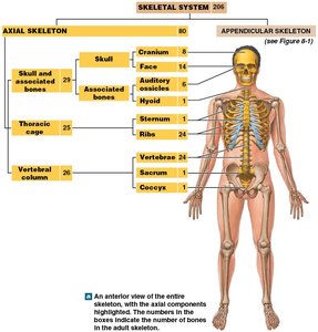
The human skeleton is divided into two main categories: the axial skeleton and the appendicular skeleton. Each division has distinct components and functions essential for support, protection, and movement.
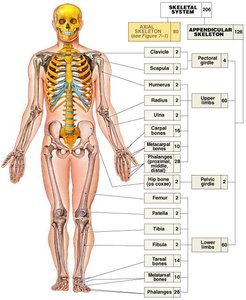
Axial Skeleton: Composed of bones of the head, neck, and trunk. It forms the longitudinal axis of the body and consists of 80 bones.
Appendicular Skeleton: Includes bones that support the limbs and their girdles, totaling 126 bones.
Functions of the Axial Skeleton
The axial skeleton serves several critical functions:
Support and Protection: Shields vital organs within body cavities, such as the brain, heart, and lungs.
Attachment Points: Provides sites for muscle attachment, facilitating movements of the head, neck, trunk, and breathing.
Stabilization: Helps stabilize parts of the appendicular skeleton.
The Skull
Cranial Bones
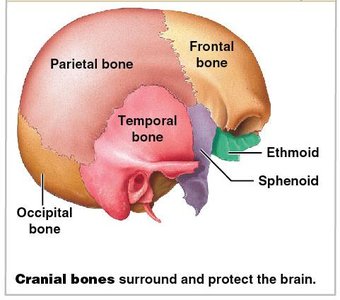
The skull contains 22 bones, divided into cranial and facial bones. The cranial bones (8 total) form the cranium, which encloses and protects the brain.
Key Cranial Bones: Frontal, parietal, temporal, occipital, sphenoid, and ethmoid.
Calvaria: The skullcap, part of the cranium.
Facial Bones
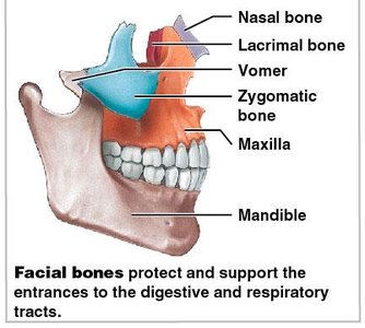
The facial bones (14 total) protect and support the entrances to the digestive and respiratory tracts.
Key Facial Bones: Nasal, lacrimal, vomer, zygomatic, maxilla, and mandible.
The Vertebral Column
Structure and Regions
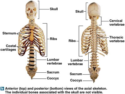
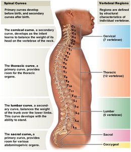
The vertebral column, or spine, protects the spinal cord and supports the head and body. It consists of 26 bones: 24 vertebrae, the sacrum, and the coccyx.
Five Regions: Cervical (7), Thoracic (12), Lumbar (5), Sacral (1), Coccygeal (1).
Spinal Curves: Primary and secondary curves develop to balance the body's weight and provide flexibility.
Vertebral Anatomy
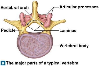
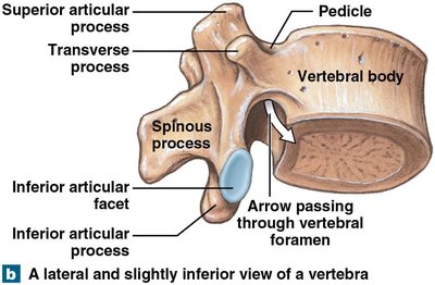
Each vertebra consists of three main parts:
Vertebral Body: The main weight-bearing portion.
Vertebral Arch: Forms the posterior part and encloses the spinal cord.
Articular Processes: Allow articulation with adjacent vertebrae.
Intervertebral Structures
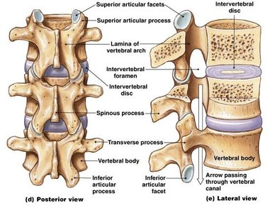
Several structures are important for vertebral function:
Intervertebral Discs: Pads of fibrocartilage separating vertebral bodies, providing cushioning and flexibility.
Intervertebral Foramina: Gaps for nerve connections to the spinal cord.
Vertebral Canal: Formed by successive vertebral foramina, enclosing the spinal cord.
The Thoracic Cage
Structure and Function
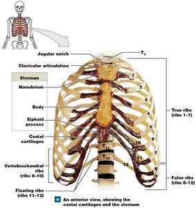
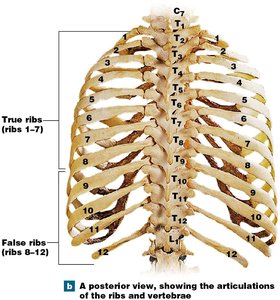
The thoracic cage is the skeleton of the chest, consisting of thoracic vertebrae, ribs, costal cartilages, and the sternum.
Functions: Protects organs of the thoracic cavity (heart, lungs, thymus), provides attachment for muscles involved in breathing, posture, and movement of pectoral girdles.
Rib Cage: Formed by ribs and sternum.
Rib Classification
There are 12 pairs of ribs, divided into two types:
True Ribs (1–7): Connected directly to the sternum by costal cartilages (vertebrosternal ribs).
False Ribs (8–12): Do not attach directly to the sternum. Ribs 8–10 are vertebrochondral (cartilages fuse and merge with rib 7), and ribs 11–12 are floating ribs (no connection to sternum).
The Appendicular Skeleton
Overview and Functions
The appendicular skeleton includes bones of the limbs and supporting girdles, accounting for 60% of the bones in the body. It enables movement and manipulation of objects.
Components: Pectoral girdle, upper limbs, pelvic girdle, lower limbs.
The Pectoral Girdle
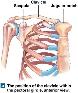
The pectoral girdle connects each arm to the body and consists of one clavicle and one scapula. It attaches to the axial skeleton only at the manubrium.
Function: Positions the shoulder joint and provides a basis for arm movement.
The Upper Limbs
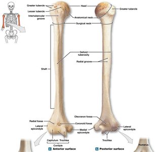
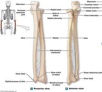
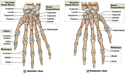
The upper limb skeleton includes the arm (humerus), forearm (ulna and radius), wrist (carpal bones), and hand (metacarpals and phalanges).
Arm: Humerus is the only bone in the brachium.
Forearm: Ulna (medial) and radius (lateral) are connected by the interosseous membrane.
Wrist and Hand: Eight carpal bones (four proximal, four distal), metacarpals, and phalanges.
The Pelvic Girdle
The pelvic girdle consists of two hip bones (coxal bones), each formed by the fusion of the ilium, ischium, and pubis. It attaches the lower limbs to the axial skeleton and is strong to bear body weight.
Function: Provides mobility and support for the lower limbs.
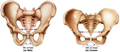
Male vs. Female Pelvis
Female Pelvis: Smoother, lighter, less prominent muscle/ligament attachments, greater angle of opening.
Male Pelvis: Heavier, more prominent attachments, smaller angle of opening.
The Lower Limbs
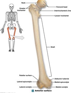
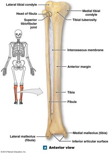
The lower limbs are specialized for weight bearing and movement. They include the femur, patella, tibia, fibula, tarsal bones, metatarsals, and phalanges.
Femur: Longest and heaviest bone, articulates with hip bone at acetabulum.
Leg: Tibia and fibula are bound together by the interosseous membrane.
Summary Table: Axial vs. Appendicular Skeleton
Division | Main Components | Number of Bones | Primary Functions |
|---|---|---|---|
Axial Skeleton | Skull, vertebral column, thoracic cage | 80 | Support, protection, muscle attachment |
Appendicular Skeleton | Pectoral girdle, upper limbs, pelvic girdle, lower limbs | 126 | Movement, manipulation, support |
Key Terms and Definitions
Axial Skeleton: The central skeleton, including the skull, vertebral column, and thoracic cage.
Appendicular Skeleton: The skeleton of the limbs and their supporting girdles.
Vertebra: A bone of the vertebral column, consisting of a body, arch, and articular processes.
Interosseous Membrane: A fibrous sheet connecting two bones, such as the radius and ulna or tibia and fibula.
Pelvic Girdle: The structure attaching the lower limbs to the axial skeleton, formed by the hip bones.
Example Application
Understanding the structure and function of the skeletal system is essential for diagnosing and treating musculoskeletal disorders, planning surgical interventions, and comprehending human movement and biomechanics.
