 Indietro
The Skeletal System: Axial Skeleton and Associated Bones
Guida di studio - Note intelligenti
 Appunti personalizzati basati sui tuoi materiali, ampliati con definizioni chiave, esempi e contesto.
The Skeletal System Overview
Axial vs. Appendicular Skeleton
The human skeleton is divided into two major parts: the axial skeleton and the appendicular skeleton. Each division contains specific bones that serve distinct structural and functional roles.
Axial Skeleton: Composed of the skull, thoracic cage, and vertebral column. It provides support and protection for the brain, spinal cord, and organs in the thorax.
Appendicular Skeleton: Includes the bones of the limbs and girdles, facilitating movement and interaction with the environment.
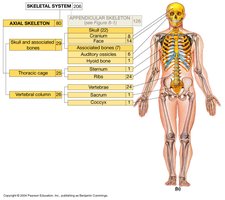
Bone Classification and Major Parts
Bones are classified based on their location and function. Each bone has major anatomical features, such as processes, foramina, and articulating surfaces, which are essential for muscle attachment and joint formation.
Axial Skeleton: Skull
Skull Structure and Components
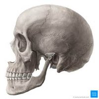
The skull consists of cranial and facial bones, as well as associated bones like the auditory ossicles and hyoid bone. The cranial bones protect the brain, while the facial bones form the structure of the face.
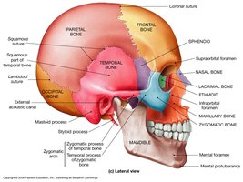
Cranial Bones (8): Occipital, Parietal (2), Frontal, Temporal (2), Sphenoid, Ethmoid
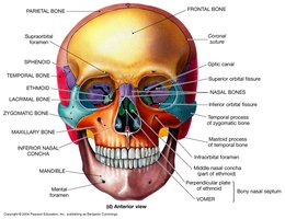
Facial Bones (14): Maxilla (2), Zygomatic (2), Palatine (2), Nasal (2), Lacrimal (2), Inferior nasal concha (2), Vomer, Mandible
Associated Bones: Auditory ossicles (6), Hyoid bone (1)
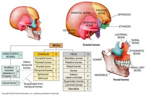
Major Parts of the Skull
Each bone of the skull has distinct anatomical landmarks:
Frontal Bone: Frontal squama, supraorbital margin, lacrimal fossa
Parietal Bones: Left and right, form the sides and roof of the cranium
Occipital Bone: Occipital condyle, external occipital crest, jugular foramen, hypoglossal canal, external occipital protuberance, internal occipital crest, foramen magnum
Temporal Bone: Squamous portion, mandibular fossa, external acoustic meatus, mastoid process, styloid process, zygomatic process, stylomastoid foramen, internal acoustic meatus, petrous portion, carotid canal
Sphenoid Bone: Sella turcica, lesser and greater wings, pterygoid process, optic canal, superior and inferior orbital fissures
Ethmoid Bone: Cribriform plate, crista galli, olfactory foramina, inferior and middle nasal concha, perpendicular plate
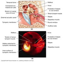
Associated Bones: Auditory Ossicles and Hyoid
Auditory Ossicles
The auditory ossicles are three small bones in each middle ear: malleus, incus, and stapes. They transmit sound vibrations from the tympanic membrane to the inner ear.
Malleus: Attached to the tympanic membrane
Incus: Intermediate bone
Stapes: Footplate fits into the oval window of the inner ear
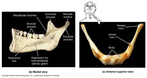
Hyoid Bone
The hyoid bone is a U-shaped bone located in the neck, not directly attached to other bones. It supports the tongue and serves as an attachment site for muscles associated with swallowing.
Skull Sutures and Sinuses
Sutures of the Cranium
Sutures are immovable joints between skull bones. Major sutures include:
Coronal Suture: Between frontal and parietal bones
Sagittal Suture: Between parietal bones
Lambdoid Suture: Between parietal and occipital bones
Squamous Suture: Between parietal and temporal bones
Facial Bones and Their Features
Key Facial Bones
The facial bones form the structure of the face and provide attachment points for muscles of facial expression and mastication.
Zygomatic Bone: Cheekbone, temporal process
Maxilla: Infraorbital foramen, alveolar process, palatine process
Palatine Bone: Forms part of the hard palate
Lacrimal Bone: Nasolacrimal canal
Nasal Bone: Bridge of the nose
Inferior Nasal Concha: Forms part of the lateral wall of the nasal cavity
Vomer: Forms part of the nasal septum
Mandible: Body, angle, ramus, condylar process, mandibular foramen, coronoid process, alveolar process, mental foramen
Thoracic Cage
Bones of the Thorax
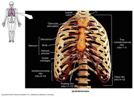
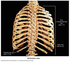
The thoracic cage protects vital organs and supports the shoulder girdles and upper limbs. It consists of the sternum, ribs, and costal cartilages.
Sternum: Jugular notch, manubrium, body, xiphoid process
Ribs:
True ribs (1-7): Attach directly to the sternum
False ribs (8-10): Attach indirectly via costal cartilage
Floating ribs (11-12): Do not attach to the sternum
Vertebral Column
Structure and Regions
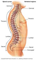
The vertebral column supports the body, protects the spinal cord, and provides attachment points for ribs and muscles. It is divided into five regions:
Cervical (7): Neck region
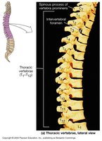
Thoracic (12): Chest region
Lumbar (5): Lower back
Sacral (5 fused): Sacrum
Coccygeal (4 fused): Coccyx
Spinal Curves and Alignment
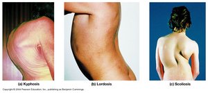
Normal spinal curves include cervical, thoracic, lumbar, and sacral. Abnormal curvatures include kyphosis, lordosis, and scoliosis.
Kyphosis: Excessive outward curvature of the thoracic region
Lordosis: Excessive inward curvature of the lumbar region
Scoliosis: Lateral curvature of the spine
Vertebrae Anatomy
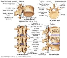
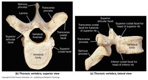
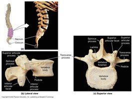
Each vertebra has a body, vertebral foramen, spinous process, transverse process, and articular facets. Intervertebral discs and foramina are important for movement and nerve passage.
Comparison of Vertebrae Types
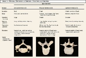
Vertebrae differ in structure and function depending on their region.
Feature | Cervical Vertebrae | Thoracic Vertebrae | Lumbar Vertebrae |
|---|---|---|---|
Location | Neck | Chest | Lower back |
Body | Small | Medium | Large |
Spinous Process | Bifid | Long, downward | Short, blunt |
Transverse Foramen | Present | Absent | Absent |
Special Features | Atlas and Axis | Costal facets | Robust structure |
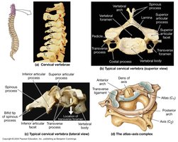
Special Vertebrae: Atlas and Axis
Cervical Vertebrae
The first two cervical vertebrae, the atlas (C1) and axis (C2), allow for head movement. The atlas supports the skull, while the axis provides a pivot for rotation.
Thoracic and Lumbar Vertebrae
Thoracic vertebrae have costal facets for rib articulation. Lumbar vertebrae are larger and support more weight.
Sacrum and Coccyx
Anatomical Features
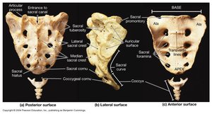
The sacrum is a triangular bone formed by the fusion of five sacral vertebrae. It articulates with the iliac bones of the pelvis. The coccyx is formed by the fusion of four small vertebrae and serves as an attachment site for ligaments and muscles.
Sacral foramina: Openings for nerves
Sacral hiatus: Terminal opening
Median sacral crest: Ridge formed by fused spinous processes
Sacral canal: Passage for spinal nerves
Sacroiliac joint: Articulation with pelvis
Auricular surface: Articulates with ilium
Sacral promontory: Anterior projection
Summary Table: Axial Skeleton Bones
Region | Bones | Number |
|---|---|---|
Skull | Cranium, Face, Associated bones | 22 |
Thoracic cage | Sternum, Ribs | 25 |
Vertebral column | Cervical, Thoracic, Lumbar, Sacrum, Coccyx | 26 |
Example: The occipital bone forms the posterior part of the skull and contains the foramen magnum, through which the spinal cord passes.
Additional info: The notes have been expanded to include definitions, anatomical context, and logical grouping for clarity and completeness.
