 Indietro
The Skeletal System: Structure, Function, and Bone Tissue
Guida di studio - Note intelligenti
 Appunti personalizzati basati sui tuoi materiali, ampliati con definizioni chiave, esempi e contesto.
The Skeletal System
Functions of the Skeletal System
The skeletal system is a complex framework that provides structural support, protection, and facilitates movement. It consists of bones, cartilages, ligaments, and other connective tissues.
Support: Provides the framework for the body, supporting soft tissues and organs.
Storage of Minerals and Lipids: Stores essential minerals such as calcium and phosphate; yellow bone marrow stores lipids.
Blood Cell Production: Red bone marrow in long bones produces red and white blood cells and platelets.
Protection: Shields vital organs such as the heart, lungs, brain, and spinal cord.
Leverage: Bones act as levers to facilitate movement by muscles.
Classification of Bones
Bone Classification by Shape
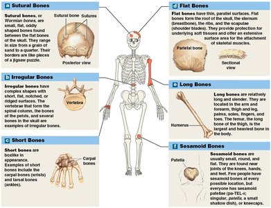
Bones are classified based on their shape and structure, which reflects their function and location in the body.
Sutural Bones: Small, flat, irregularly shaped bones found between the flat bones of the skull.
Flat Bones: Thin bones with parallel surfaces, such as the sternum, ribs, and skull bones. They provide protection and extensive surface area for muscle attachment.
Irregular Bones: Complex shapes with short, flat, notched, or ridged surfaces, such as vertebrae and pelvic bones.
Short Bones: Small and boxy, found in the wrist (carpal bones) and ankle (tarsal bones).
Long Bones: Long and slender, found in the arms, legs, fingers, and toes. They are crucial for movement and leverage.
Sesamoid Bones: Small, round, and flat bones found within tendons, such as the patella.
Bone Markings
Bones display surface features known as bone markings, which include projections (for muscle and ligament attachment) and openings or depressions (for passage of blood vessels and nerves).
Structure of Bones
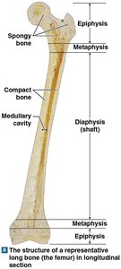
Structure of a Long Bone
Long bones have a distinct anatomy that supports their function in movement and weight-bearing.
Diaphysis: The shaft, composed of compact bone surrounding the medullary (marrow) cavity.
Epiphysis: The expanded ends, primarily made of spongy bone.
Metaphysis: The region where the diaphysis and epiphysis meet.
Structure of Flat Bones
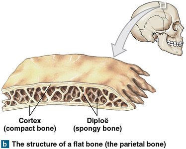
Flat bones, such as the parietal bones of the skull, consist of spongy bone sandwiched between two layers of compact bone. The spongy bone layer within the cranium is called the diploë.
Bone Tissue
Characteristics of Bone Tissue
Bone tissue is a dense, supportive connective tissue with a solid matrix composed of collagen fibers and calcium salts. It contains specialized cells and is organized to facilitate nutrient exchange and repair.
Dense Matrix: Due to calcium salt deposits.
Osteocytes: Bone cells housed in lacunae, organized around blood vessels.
Canaliculi: Narrow passageways for exchange of nutrients, wastes, and gases.
Periosteum: A membrane covering the outer surface of bones, except at joints, consisting of fibrous and cellular layers.
Types of Bone Cells
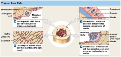
Bone tissue contains four main cell types, each with distinct functions in bone formation, maintenance, and remodeling.
Osteogenic Cells: Stem cells that divide to produce osteoblasts; located in the periosteum and endosteum; assist in fracture repair.
Osteoblasts: Immature cells that synthesize new bone matrix during osteogenesis; become osteocytes when surrounded by matrix.
Osteocytes: Mature bone cells that maintain the bone matrix and assist in repair; reside in lacunae and communicate via canaliculi.
Osteoclasts: Multinucleate cells that dissolve bone matrix by secreting acids and enzymes; important for mineral homeostasis.
Bone Cell Life Cycle
Bone cell development follows a cycl e: osteoprogenitor cells differentiate into osteoblasts, which build bone and mature into osteocytes. Osteoclasts, derived from a different lineage, resorb bone matrix.
Compact Bone and Spongy Bone
Structure of Compact Bone
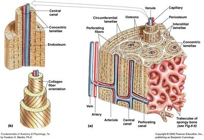
Compact bone is organized into osteons, the functional unit, which are concentric rings of lamellae surrounding a central canal containing blood vessels.
Osteon: Basic unit of mature compact bone.
Lamellae: Layers of bone matrix; concentric, interstitial, and circumferential types.
Central Canal: Contains blood vessels and nerves.
Perforating Canals: Carry blood vessels into deep bone and marrow.
Structure of Spongy Bone
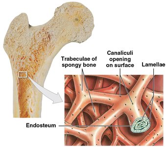
Spongy bone lacks osteons and consists of a network of trabeculae. Spaces between trabeculae are filled with red bone marrow, which forms blood cells, and yellow bone marrow, which stores fat.
Bone Membranes
Periosteum
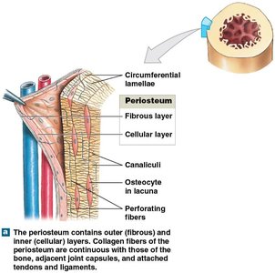
The periosteum is a membrane covering the external surface of bones, except at joints. It consists of an outer fibrous layer and an inner cellular layer, and its fibers are interwoven with those of tendons.
Functions: Isolates bone from surrounding tissues, provides a route for blood vessels and nerves, and participates in bone growth and repair.
Perforating Fibers: Collagen fibers that anchor the periosteum to bone tissue, increasing attachment strength.
Endosteum
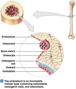
The endosteum is an incomplete cellular layer lining the medullary cavity, covering trabeculae of spongy bone, and lining central canals of compact bone. It is active during bone growth, repair, and remodeling.
Cell Types: Contains osteogenic cells, osteoblasts, and osteoclasts.
Bone Formation and Growth
Ossification and Remodeling
Bone formation (ossification or osteogenesis) involves the deposition of calcium salts (calcification) and continuous remodeling. Remodeling can alter the matrix or the shape and internal structure of bone.
Osteocytes: Continuously remove and replace calcium salts.
Osteoblasts and Osteoclasts: Work in balance to build and resorb bone.
Exercise, Nutrition, and Hormones
Effects of Exercise on Bone
Exercise stimulates mineral recycling and osteoblast activity, making bones thicker and stronger. Weight-bearing exercise is particularly effective.
Bone Degeneration: Bone mass can decrease rapidly with inactivity, losing up to one-third in a few weeks.
Summary Table: Types of Bone Cells
Cell Type | Function | Location |
|---|---|---|
Osteogenic Cell | Stem cell; produces osteoblasts | Periosteum, endosteum |
Osteoblast | Builds new bone matrix | Bone surface |
Osteocyte | Maintains bone matrix | Lacunae within bone |
Osteoclast | Resorbs bone matrix | Bone surface, endosteum |
Summary Table: Bone Types by Shape
Bone Type | Example | Function |
|---|---|---|
Sutural | Skull sutures | Fill gaps between flat bones |
Flat | Parietal bone, sternum | Protection, muscle attachment |
Irregular | Vertebrae | Complex shape, support |
Short | Carpal bones | Support, limited movement |
Long | Femur, humerus | Leverage, movement |
Sesamoid | Patella | Protect tendons |
