 Indietro
The Skeleton: Structure, Divisions, and Clinical Relevance
Guida di studio - Note intelligenti
 Appunti personalizzati basati sui tuoi materiali, ampliati con definizioni chiave, esempi e contesto.
The Skeleton
Overview of the Skeletal System
The human skeleton is a complex framework of bones, cartilages, joints, and ligaments that provides structural support, protection, and movement. It accounts for approximately 20% of total body mass and is divided into two main divisions: the axial skeleton and the appendicular skeleton.
Axial Skeleton: Forms the longitudinal axis of the body, supporting the head, neck, and trunk, and protecting the brain, spinal cord, and thoracic organs.
Appendicular Skeleton: Comprises the bones of the limbs and their girdles, facilitating movement and interaction with the environment.
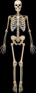
Main Divisions of the Skeleton
Axial Skeleton: 80 bones divided into the skull, vertebral column, and thoracic cage.
Appendicular Skeleton: 126 bones including the pectoral girdle, pelvic girdle, and bones of the upper and lower limbs.
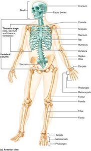
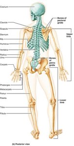
Axial Skeleton
Skull: Cranial and Facial Bones
The skull is the most complex bony structure, formed by cranial and facial bones. Cranial bones enclose the brain and provide attachment sites for head and neck muscles, while facial bones form the framework of the face and house special sense organs.
Cranial Bones (8): Frontal, parietal (2), occipital, temporal (2), sphenoid, ethmoid.
Facial Bones (14): Mandible, maxillae (2), zygomatic (2), nasal (2), lacrimal (2), palatine (2), inferior nasal conchae (2), vomer.
Sutures: Immovable joints connecting skull bones, with a serrated appearance.
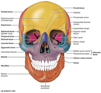
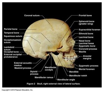
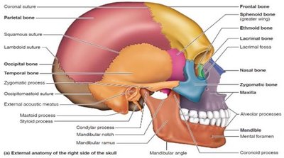
Major Markings and Cavities of the Skull
Foramina, Canals, Fissures: Openings for passage of nerves and blood vessels.
Cranial Fossae: Depressions in the cranial base that accommodate the brain's lobes.
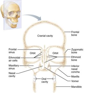
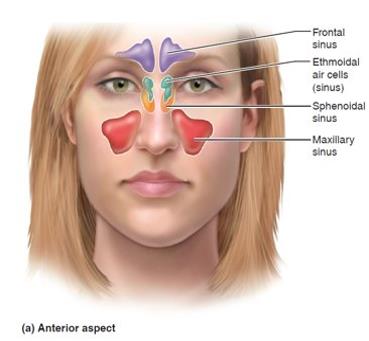
Sinuses: Air-filled spaces that lighten the skull and enhance voice resonance.
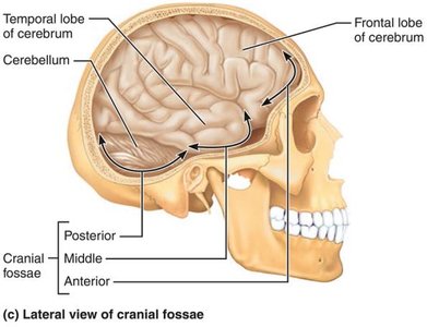
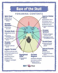
Fontanels and Sutures in Infants
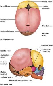
Infant skulls have more bones than adult skulls, with unfused regions called fontanels ('soft spots'). These allow for brain growth and ease of birth. Sutures are flexible joints that ossify with age.
Vertebral Column
Structure and Regions
The vertebral column, or spine, extends from the skull to the pelvis and consists of 26 irregular bones in five major regions. It transmits body weight, protects the spinal cord, and provides attachment points for ribs and muscles.
Cervical (7 vertebrae)
Thoracic (12 vertebrae)
Lumbar (5 vertebrae)
Sacrum (5 fused vertebrae)
Coccyx (3-5 fused vertebrae)
Curvatures and Ligaments
The vertebral column has four main curvatures that increase resilience and flexibility. Ligaments and trunk muscles support the column.
Cervical and lumbar: Concave posteriorly
Thoracic and sacral: Convex posteriorly
Intervertebral Discs and Clinical Implications
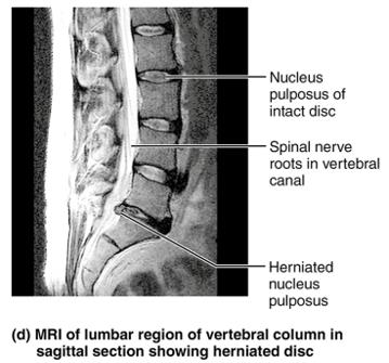
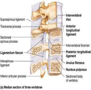
Intervertebral discs act as shock absorbers and consist of a gelatinous nucleus pulposus and a collagenous anulus fibrosus. Trauma can cause herniation, leading to nerve compression and pain.
Abnormal Spinal Curvatures
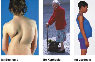
Scoliosis: Lateral curvature, often in thoracic region.
Kyphosis: Dorsal thoracic curvature (hunchback).
Lordosis: Accentuated lumbar curvature (swayback).
Thoracic Cage
Components and Functions
The thoracic cage consists of the thoracic vertebrae, sternum, costal cartilages, and ribs. It protects vital organs, supports the shoulder girdles, and provides attachment sites for muscles.
Sternum: Manubrium, body, and xiphoid process.
Ribs: 12 pairs; true ribs (1–7), false ribs (8–10), and floating ribs (11–12).
Appendicular Skeleton
Pectoral Girdle and Upper Limb
The pectoral girdle consists of the clavicles and scapulae, attaching the upper limbs to the trunk and allowing a wide range of motion. The upper limb includes the humerus, radius, ulna, carpals, metacarpals, and phalanges.
Humerus: Only bone of the arm, articulates with scapula, radius, and ulna.
Radius and Ulna: Form the forearm, connected by an interosseous membrane.
Hand: 8 carpals, 5 metacarpals, 14 phalanges.
Pelvic Girdle and Lower Limb
The pelvic girdle is formed by two hip bones and the sacrum, transmitting body weight to the lower limbs and supporting pelvic organs. The lower limb includes the femur, patella, tibia, fibula, tarsals, metatarsals, and phalanges.
Femur: Largest and strongest bone, articulates with the acetabulum and tibia.
Tibia and Fibula: Form the leg, connected by an interosseous membrane.
Foot: 7 tarsals, 5 metatarsals, 14 phalanges.
Sexual Dimorphism in the Skeleton
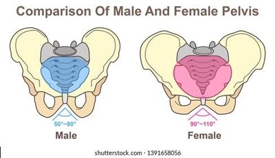
There are notable differences between male and female skeletons, especially in the pelvis, which is adapted in females for childbirth.
Characteristic | Female | Male |
|---|---|---|
General structure | Tilted forward; adapted for childbearing; broad, shallow true pelvis | Tilted less far forward; adapted for support; narrow, deep true pelvis |
Bone thickness | Lighter, thinner, smoother | Heavier, thicker, more prominent markings |
Acetabula | Smaller, farther apart | Larger, closer together |
Pubic arch angle | Broader (80–90°) | More acute (50–60°) |
Developmental Aspects and Clinical Implications
Primary curvatures: Thoracic and sacral, present at birth (C-shaped spine).
Secondary curvatures: Cervical and lumbar, develop as child grows.
Aging: Intervertebral discs lose hydration and elasticity, increasing risk of herniation and height loss.
Developmental Dysplasia of the Hip (DDH): Partial or complete dislocation of the femoral head, more common in breech births.
