 Indietro
The Skeleton: Structure, Regions, and Key Features
Guida di studio - Note intelligenti
 Appunti personalizzati basati sui tuoi materiali, ampliati con definizioni chiave, esempi e contesto.
The Skeleton: Structure, Regions, and Key Features
Introduction to the Skeleton
The human skeleton is a complex framework of bones that supports and protects the body, facilitates movement, and houses critical organs. It is divided into two main divisions: the axial skeleton and the appendicular skeleton.
The Axial Skeleton
Overview and Major Functions
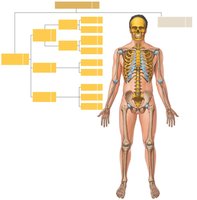
Axial Skeleton: Forms the longitudinal axis of the body and consists of 80 bones, including the skull, vertebral column, and thoracic cage.
Functions:
Supports and protects the brain, spinal cord, and organs in the ventral cavity.
Provides attachment points for muscles that adjust the head, neck, and trunk, perform respiratory movements, and stabilize the appendicular skeleton.
Bones of the Skull
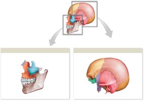
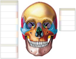
The skull is composed of cranial and facial bones, which protect the brain and form the structure of the face.
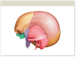
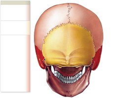
Cranial Bones (8): Frontal, occipital, sphenoid, ethmoid, 2 parietal, 2 temporal.
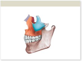
Facial Bones (14): 2 zygomatic, 2 maxilla, 2 lacrimal, 2 nasal, 2 palatine, vomer, 2 inferior nasal concha, mandible.
Associated Bones: 3 pairs of auditory ossicles and the hyoid bone.
Skull Sutures and Markings
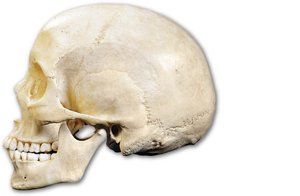
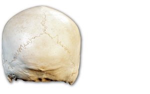
Sutures are immovable joints between skull bones. Key markings include:
Coronal Suture: Between frontal and parietal bones.
Squamous Suture: Between parietal and temporal bones.
Lambdoid Suture: Between parietal and occipital bones.
Sagittal Suture: Between the two parietal bones.
Key Features of the Skull
Inferior Nasal Conchae: Create air turbulence and warm/humidify air.
External Occipital Crest: Attachment for ligament stabilizing neck vertebrae.
Mastoid Process: Muscle attachment for head rotation.
Styloid Process: Suspends hyoid bone and serves as muscle attachment.
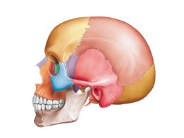
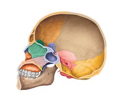
Lateral and Sagittal Views of the Skull
Alveolar Processes: Sockets for teeth in maxillae and mandible.
Mental Protuberance: Chin muscle attachment.
Mandibular Angle: Posterior, inferior corner of the jaw.
Zygomatic Arch: Formed by temporal and zygomatic bones.
External Acoustic Meatus: Opening for sound waves to reach the tympanic membrane.
Frontal Sinuses: Variable in size, appear around age 6.
Petrous Part of Temporal Bone: Encloses inner ear structures.
Internal Acoustic Meatus: Passage for blood vessels and nerves to the inner ear.
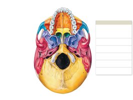
Inferior and Internal Views of the Skull
Mandibular Fossa: Articulates with the mandible.
Occipital Condyles: Articulate with the first cervical vertebra.
Foramen Magnum: Connects cranial cavity with vertebral canal.
Carotid Canal: Passage for the internal carotid artery.
Jugular Foramen: Passage for the jugular vein.
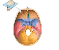
Sphenoid, Ethmoid, and Palatine Bones
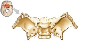
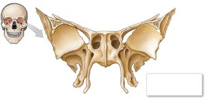
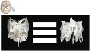
Sphenoid Bone: Forms part of the cranial floor, unites cranial and facial bones, contains optic canals, sella turcica, and sphenoidal sinuses.
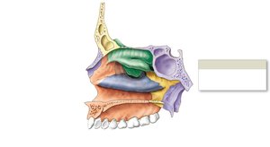
Ethmoid Bone: Forms part of the anterior cranial floor, roof of nasal cavity, and nasal septum. Contains cribriform plate (olfactory nerves), crista galli, ethmoidal labyrinth, and nasal conchae.
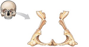
Palatine Bones: Form the posterior hard palate and contribute to the floor of the orbit.
Orbital and Nasal Complexes
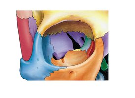
Orbital Complex: Seven bones form the orbit, protecting the eyes and associated structures.
Lacrimal Sulcus: Groove for tear drainage.
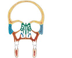
Paranasal Sinuses: Air-filled chambers in frontal, sphenoid, ethmoid, palatine, and maxillae bones; lighten the skull, resonate voice, and provide mucous membrane surface.
Mandible and Associated Bones
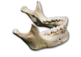
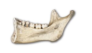
Mandible: Lower jaw, supports lower teeth, articulates with temporal bone at the temporomandibular joint.
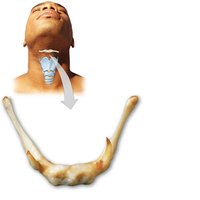
Hyoid Bone: U-shaped bone in the neck, not attached to other bones, supports muscles of the larynx, pharynx, and tongue.
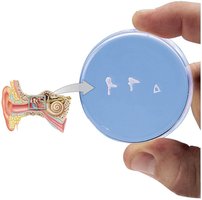
Auditory Ossicles: Malleus, incus, and stapes; conduct sound vibrations in the middle ear.
Developmental Differences in the Skull
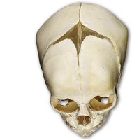
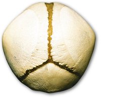
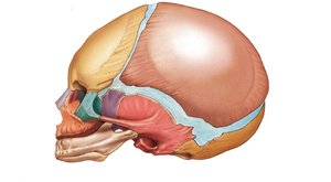
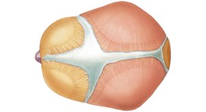
Fontanelles: Fibrous areas between cranial bones in infants, allowing for brain growth and skull flexibility during birth. The anterior fontanelle is the largest and persists until about age 2.
Skull Growth: Most significant before age 5, after which cranial sutures ossify.
The Vertebral Column
Structure and Regions
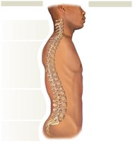
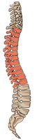
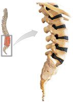
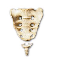
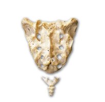
Vertebral Column: 26 bones (24 vertebrae, sacrum, coccyx); supports head, neck, trunk, protects spinal cord, and transfers weight to appendicular skeleton.
Regions:
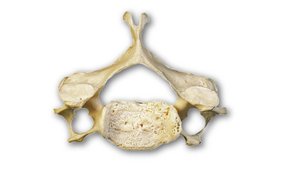
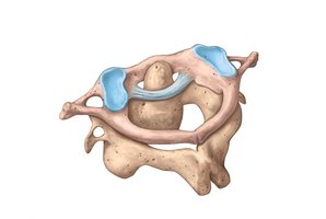
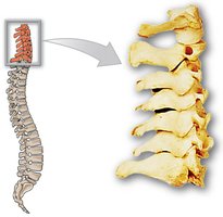
Cervical (7 vertebrae): Supports the head.
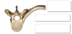
Thoracic (12 vertebrae): Articulates with ribs.
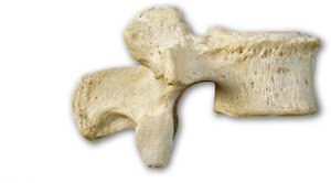
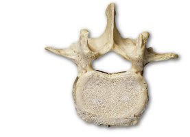
Lumbar (5 vertebrae): Largest, supports most weight.
Sacrum (5 fused vertebrae): Foundation for pelvic girdle.
Coccyx (3-5 fused vertebrae): Tailbone.
Parts of a Vertebra
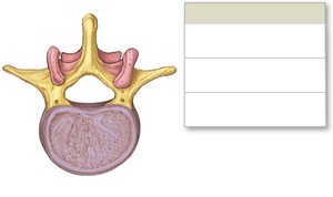
Vertebral Foramen: Passage for the spinal cord.
Intervertebral Foramen: Openings for spinal nerves.
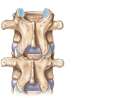
Articular Processes: Form joints with adjacent vertebrae.
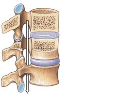
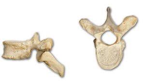
Vertebral Body: Weight-bearing portion.
Intervertebral Disk: Cartilaginous pad absorbing shock and permitting movement.
Pedicles and Laminae: Form the vertebral arch.
Transverse and Spinous Processes: Muscle and ligament attachment points.
Regional Comparison of Vertebrae
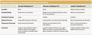
Feature | Cervical Vertebrae (7) | Thoracic Vertebrae (12) | Lumbar Vertebrae (5) |
|---|---|---|---|
Location | Neck | Chest | Inferior portion of back |
Vertebral Body | Small, oval, curved faces | Larger, heart-shaped; flat faces for rib articulations | Massive, oval faces |
Vertebral Foramen | Large | Smaller | Smallest |
Spinous Process | Long, split tip; points inferiorly | Long, slender; not split; points inferiorly | Blunt, broad; points posteriorly |
Transverse Processes | Have transverse foramina | All but two have facets for rib articulations | Short; no articular facets or transverse foramina |
Functions | Support skull, stabilize relative positions of brain and spinal cord, allow controlled head movement | Support weight of head, neck, upper limbs, chest; articulate with ribs to allow changes in volume of thoracic cage | Support weight of head, neck, upper limbs, trunk |
The Thoracic Cage
Structure and Functions
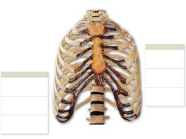
Thoracic Cage: Composed of the sternum, ribs, costal cartilages, and thoracic vertebrae; protects thoracic organs and supports respiration.
Sternum: Manubrium (articulates with clavicle and rib 1), body (articulates with ribs 2-7), xiphoid process (muscle attachment).
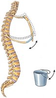
Ribs: 12 pairs; true ribs (1-7, direct attachment), false ribs (8-10, indirect attachment), floating ribs (11-12, no attachment to sternum).
The Appendicular Skeleton
Overview
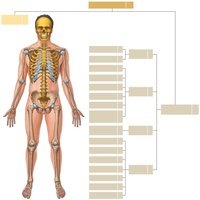
Appendicular Skeleton: Includes the bones of the limbs and girdles (pectoral and pelvic) that attach them to the axial skeleton. Contains 126 bones.
Pectoral Girdle
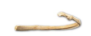
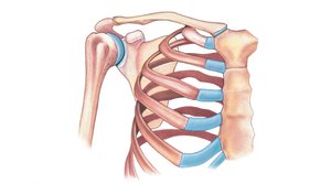
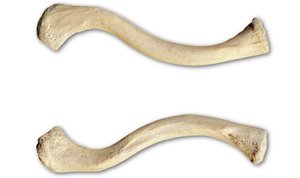
Clavicle: Sternal end articulates with sternum; acromial end with scapula.
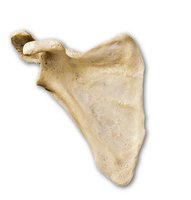
Scapula: Acromion (articulates with clavicle), glenoid cavity (articulates with humerus), scapular spine (muscle attachment).
Arm and Forearm Bones
Humerus: Head articulates with scapula; surgical neck is a common fracture site; epicondyles for muscle attachment; olecranon, radial, and coronoid fossae for articulation with ulna and radius.
Radius: Lateral bone; radial tuberosity for biceps attachment; styloid process stabilizes wrist.
Ulna: Medial bone; olecranon forms elbow; trochlear notch articulates with humerus; ulnar head articulates with wrist.
Wrist and Hand Bones
Carpal Bones: Eight bones in two rows (scaphoid, lunate, triquetrum, pisiform, trapezium, trapezoid, capitate, hamate).
Metacarpals: Five bones supporting the hand.
Phalanges: 14 bones per hand; thumb (pollex) has 2, other fingers have 3 each.
Pelvic Girdle and Pelvis
Pelvic Girdle: Two coxal bones, each formed by fusion of ilium, ischium, and pubis.
Acetabulum: Socket for femur head.
Greater Sciatic Notch: Passage for sciatic nerve.
Pubic Symphysis: Cartilaginous joint between pubic bones.
Obturator Foramen: Large opening for nerve and muscle passage.
Pelvic Differences: Male vs. Female
Female Pelvis: Broader, shallower, wider pelvic inlet and outlet, less curvature of sacrum and coccyx, adapted for childbirth.
Male Pelvis: Narrower, deeper, more curved sacrum and coccyx.
Thigh and Leg Bones
Femur: Longest and strongest bone; neck, greater trochanter, and linea aspera are key features.
Patella: Kneecap; attachment for quadriceps tendon and patellar ligament.
Tibia: Larger, medial lower leg bone; tibial tuberosity for ligament attachment; medial malleolus forms ankle prominence.
Fibula: Thinner, lateral bone; lateral malleolus forms ankle prominence; does not bear weight.
Ankle and Foot Bones
Tarsal Bones: Seven bones; calcaneus (heel) and talus are most significant for weight transmission.
Metatarsals: Five bones forming the foot's distal portion.
Phalanges: 14 bones per foot; hallux (big toe) has 2, others have 3 each.
Arches of the Foot: Longitudinal and transverse arches distribute body weight and provide support.
Medical Terminology Associated with the Axial Skeleton
Laminectomy: Surgical removal of a vertebral lamina, often to relieve herniated disc symptoms.
Spinal Fusion: Stabilization of vertebrae using bone grafts or synthetic devices.
Whiplash: Neck injury from rapid hyperextension and hyperflexion, common in car accidents.
Clubfoot (Talipes Equinovarus): Inherited deformity with twisted foot and increased arch angle.
Genu Valgum: Knock-knee deformity; knees close together, ankles apart.
Genu Varum: Bowleg deformity; knees apart, lower limbs bowed medially.
