 Indietro
The Special Senses: Structure and Function of the Eye
Guida di studio - Note intelligenti
 Appunti personalizzati basati sui tuoi materiali, ampliati con definizioni chiave, esempi e contesto.
Special Senses: The Eye
Overview of the Eyeball Structure
The human eye is a complex sensory organ responsible for vision. Its structure is specialized to capture light, focus it, and convert it into neural signals for the brain to interpret. The eyeball consists of three main layers, internal cavities filled with fluids, and various specialized structures.
Fibrous Layer: Outermost layer, includes the sclera and cornea.
Vascular Layer: Middle pigmented layer, includes the choroid, ciliary body, and iris.
Inner Layer (Retina): Contains photoreceptors and neurons for visual processing.
Internal Cavities: Filled with aqueous and vitreous humors, separated by the lens into anterior and posterior segments.
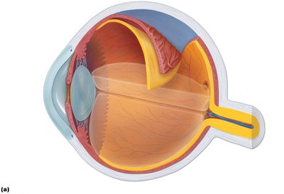
Fibrous Layer
The fibrous layer forms the outermost protective coat of the eye and is composed of dense avascular connective tissue. It consists of two regions:
Sclera: The opaque, white posterior region that protects and shapes the eyeball.
Cornea: The transparent anterior region that allows light to enter and bends (refracts) light as it enters the eye.
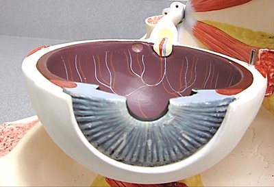
Vascular Layer
The vascular layer is the middle pigmented layer of the eye, providing nourishment and controlling lens shape and pupil size. It includes:
Choroid: Supplies blood to all layers of the eyeball.
Ciliary Body: Contains smooth muscle (ciliary muscles) that control lens shape; ciliary zonules (suspensory ligaments) attach the lens.
Iris: The colored part of the eye, continuous with the ciliary body, containing the pupil (central opening) that regulates the amount of light entering the eye.
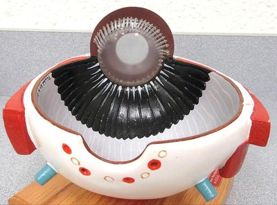
Inner Layer (Retina)
The retina is the innermost layer of the eye, responsible for converting light into neural signals. It consists of two layers:
Pigmented Layer: Absorbs light and prevents scattering.
Neural Layer: Contains three main types of neurons: photoreceptors (rods and cones), bipolar cells, and ganglion cells.
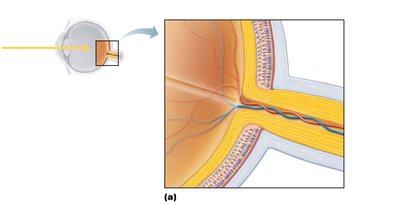
Photoreceptors: Rods and Cones
Rods: Specialized for dim light and peripheral vision; more numerous and sensitive to light but do not provide color vision or sharp images.
Cones: Responsible for bright light and high-resolution color vision; concentrated in the macula lutea and especially in the fovea centralis, the region of highest visual acuity.
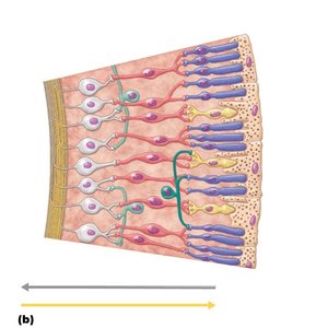
Histology of the Retina
The retina's layered structure can be observed under the microscope, showing the arrangement of photoreceptors, bipolar cells, and ganglion cells.
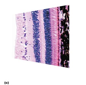
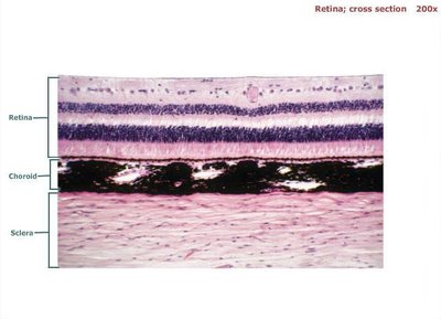
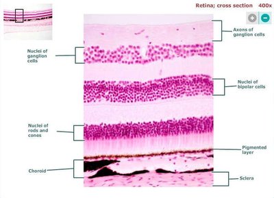
Key Structures of the Eye
Optic Nerve: Transmits visual information from the retina to the brain.
Optic Disc (Blind Spot): Site where the optic nerve exits the eye; lacks photoreceptors.
Fovea Centralis: Small pit in the macula lutea with the highest density of cones and best visual acuity.
Lens: Transparent structure that focuses light onto the retina; its shape is adjusted by the ciliary muscles.
Aqueous Humor: Clear fluid in the anterior segment, providing nutrients and maintaining intraocular pressure.
Vitreous Humor: Gel-like substance in the posterior segment, maintaining eye shape and holding the retina in place.
Extrinsic Eye Muscles
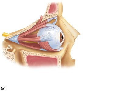
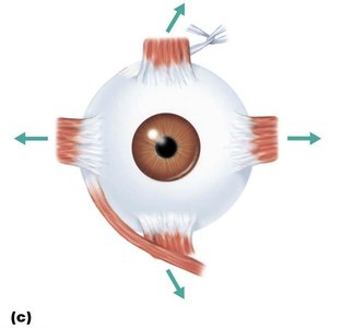
Six extrinsic eye muscles control the movement of the eyeball, allowing it to follow moving objects, maintain its shape, and hold it in the orbit. These muscles are innervated by cranial nerves III (oculomotor), IV (trochlear), and VI (abducens).
Muscle | Action | Controlling Cranial Nerve |
|---|---|---|
Lateral rectus | Moves eye laterally | VI (abducens) |
Medial rectus | Moves eye medially | III (oculomotor) |
Superior rectus | Elevates eye and turns it medially | III (oculomotor) |
Inferior rectus | Depresses eye and turns it medially | III (oculomotor) |
Inferior oblique | Elevates eye and turns it laterally | III (oculomotor) |
Superior oblique | Depresses eye and turns it laterally | IV (trochlear) |
Lacrimal Apparatus
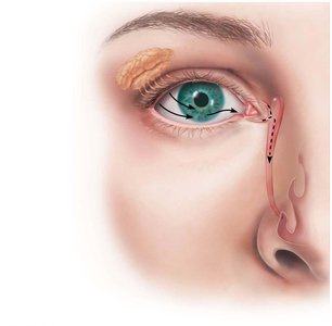
The lacrimal apparatus produces, distributes, and removes tears. Tears lubricate the eye, provide nutrients, and contain enzymes that protect against infection. The apparatus includes the lacrimal gland, ducts, sac, and nasolacrimal duct.
Summary Table: Main Structures of the Eye
Structure | Function |
|---|---|
Sclera | Protects and shapes the eyeball |
Cornea | Refracts light entering the eye |
Choroid | Provides blood supply to the eye |
Ciliary Body | Controls lens shape |
Iris | Regulates pupil size and light entry |
Retina | Contains photoreceptors for vision |
Lens | Focuses light onto the retina |
Optic Nerve | Transmits visual signals to the brain |
Additional info:
The tapetum lucidum is a reflective layer found in some animals (not humans) that enhances night vision by reflecting light back through the retina.
Dissection images (not shown here) can help visualize the anatomical relationships between these structures in real tissue.
