 Indietro
The Urinary System: Structure and Function of the Kidney and Associated Organs
Guida di studio - Note intelligenti
 Appunti personalizzati basati sui tuoi materiali, ampliati con definizioni chiave, esempi e contesto.
The Urinary System
Overview of the Urinary System
The urinary system is responsible for filtering blood, removing waste products, and regulating fluid and electrolyte balance in the body. Its main organs include the kidneys, ureters, urinary bladder, and urethra. The kidneys play a central role in homeostasis by forming urine and regulating blood pressure, pH, and electrolyte levels.
The Kidney
Gross Anatomy of the Kidney
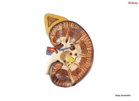
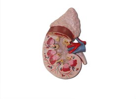
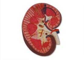
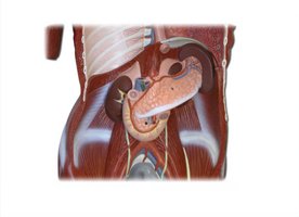
The kidneys are paired, bean-shaped organs located retroperitoneally on either side of the vertebral column. Each kidney is surrounded by a fibrous capsule and contains distinct regions: the cortex, medulla, and renal pelvis.
Renal Cortex: The outer region containing the majority of nephrons.
Renal Medulla: The inner region composed of renal pyramids, which contain the collecting ducts and loops of Henle.
Renal Pelvis: The funnel-shaped structure that collects urine from the major calyces and channels it into the ureter.
Renal Capsule: A tough, fibrous layer surrounding the kidney for protection.
Renal Columns: Extensions of cortical tissue between the renal pyramids.
Renal Papillae: The tips of the pyramids where urine is discharged into the minor calyces.
Major and Minor Calyces: Chambers that collect urine from the papillae and funnel it toward the renal pelvis.
Renal Artery and Vein: Blood vessels supplying and draining the kidney.
Ureter: Tube that carries urine from the renal pelvis to the urinary bladder.
Associated Structures
Adrenal Gland: Sits atop each kidney and secretes hormones such as adrenaline and cortisol.
Suprarenal Artery and Vein: Vessels supplying and draining the adrenal gland.
Microscopic Structure: The Nephron
Nephron Anatomy and Types
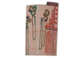
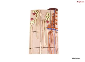
The nephron is the functional unit of the kidney, responsible for filtering blood and forming urine. Each kidney contains about one million nephrons, which are classified as cortical or juxtamedullary based on their location and structure.
Cortical Nephrons: Located primarily in the renal cortex; have short loops of Henle.
Juxtamedullary Nephrons: Located near the cortex-medulla junction; have long loops of Henle that extend deep into the medulla, crucial for concentrating urine.
Renal Corpuscle
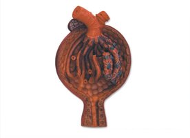
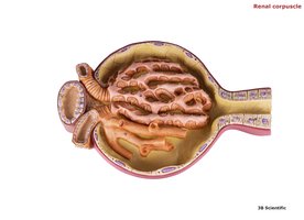
The renal corpuscle is the initial filtering component of the nephron, consisting of the glomerulus and the glomerular (Bowman's) capsule.
Glomerulus: A tuft of capillaries where blood filtration occurs.
Glomerular Capsule: A double-walled epithelial cup that surrounds the glomerulus and collects the filtrate.
Afferent Arteriole: Brings blood into the glomerulus.
Efferent Arteriole: Carries blood away from the glomerulus.
Podocytes: Specialized cells in the inner layer of the capsule that aid in filtration.
Nephron Tubule Segments
Proximal Convoluted Tubule (PCT): Site of most reabsorption of water, ions, and nutrients.
Loop of Henle (Nephron Loop): Descending and ascending limbs that concentrate urine by creating a gradient in the medulla.
Distal Convoluted Tubule (DCT): Further adjusts the composition of urine; site of hormone action (e.g., aldosterone, ADH).
Collecting Duct: Receives urine from multiple nephrons and channels it to the renal pelvis.
Blood Supply of the Nephron
Cortical Radiate Vessels: Supply blood to the cortex and nephrons.
Arcuate Vessels: Arch over the base of the pyramids, connecting interlobar and cortical radiate arteries.
Vasa Recta: Capillary network surrounding the loop of Henle in juxtamedullary nephrons, important for urine concentration.
Urinary Tract and Associated Structures
Ureter
The ureter is a muscular tube that transports urine from the renal pelvis to the urinary bladder via peristaltic contractions.
Urinary Bladder
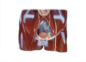
The urinary bladder is a hollow, muscular organ that stores urine until micturition (urination). It is lined by transitional epithelium and contains a region called the trigone, defined by the openings of the ureters and the urethra.
Urethra
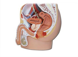
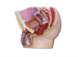
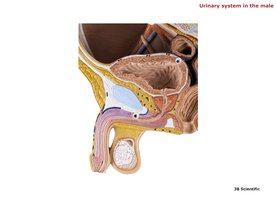
The urethra is the tube that conveys urine from the bladder to the exterior of the body. Its length and structure differ between males and females.
Male Urethra: Divided into prostatic, membranous (intermediate), and spongy (penile) segments.
Female Urethra: Shorter and opens anterior to the vaginal opening.
Sex Differences in the Urinary System
Male: Urethra passes through the prostate and penis; longer and serves both urinary and reproductive functions.
Female: Urethra is shorter and separate from the reproductive tract.
Relevant Images of the Urinary Tract
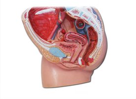
Summary Table: Main Structures of the Kidney
Structure | Location | Function |
|---|---|---|
Renal Cortex | Outer region of kidney | Contains renal corpuscles and most nephrons |
Renal Medulla | Inner region, contains pyramids | Contains loops of Henle and collecting ducts |
Renal Pelvis | Central cavity | Collects urine for passage to ureter |
Nephron | Spans cortex and medulla | Filters blood, forms urine |
Ureter | From renal pelvis to bladder | Transports urine |
Urinary Bladder | Pelvic cavity | Stores urine |
Urethra | From bladder to exterior | Expels urine from body |
Key Equations
Glomerular Filtration Rate (GFR):
Renal Clearance:
Where: C = clearance rate U = concentration of substance in urine V = urine flow rate P = concentration of substance in plasma
Additional info: The above notes integrate anatomical and physiological details relevant to the urinary system, focusing on the kidney and its microscopic and macroscopic structures, as well as the urinary tract in both sexes. Images are included only where they directly reinforce the described anatomical features.
