 Indietro
The Urinary System: Structure and Function
Guida di studio - Note intelligenti
 Appunti personalizzati basati sui tuoi materiali, ampliati con definizioni chiave, esempi e contesto.
The Urinary System
Overview and Functions
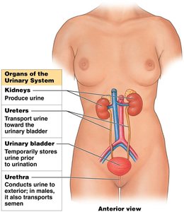
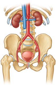
The urinary system is essential for removing metabolic wastes, regulating blood composition, and maintaining homeostasis. It consists of paired kidneys, ureters, a urinary bladder, and a urethra. Each organ plays a distinct role in urine production, transport, storage, and elimination.
Excretion: Removal of metabolic wastes from body fluids.
Elimination: Discharge of wastes from the body.
Homeostatic Regulation: Maintains blood volume, solute concentration, and pH.
Homeostatic Functions
Regulation of Blood Volume and Pressure: Adjusts water loss in urine, influenced by endocrine signals.
Regulation of Plasma Ion Concentrations: Controls sodium, potassium, chloride, and other ions lost in urine.
Stabilization of Blood pH: Manages hydrogen and bicarbonate ion loss.
Conservation of Nutrients: Prevents loss of valuable nutrients while removing wastes.
Assistance to Liver: Detoxifies poisons and deaminates amino acids during starvation.
Kidneys
Location and Structure
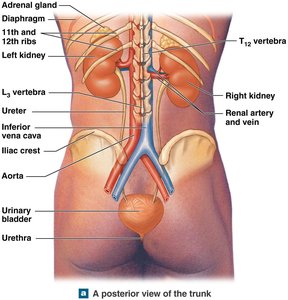
The kidneys are retroperitoneal organs located on either side of the vertebral column. The left kidney is slightly superior to the right, and each is capped by an adrenal gland. Their position is stabilized by the peritoneum, adjacent organs, and connective tissues.
Internal Structure
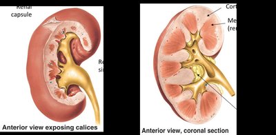
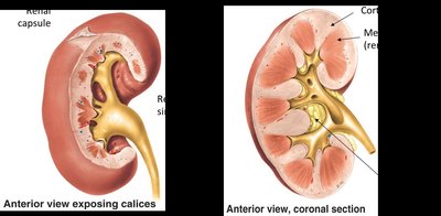
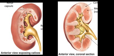
Renal Cortex: The outer, granular region in contact with the fibrous capsule.
Renal Medulla: Contains 6–18 renal pyramids, each with a base abutting the cortex and a tip (renal papilla) projecting into the renal sinus.
Renal Columns: Bands of cortical tissue separating adjacent pyramids, extending into the medulla.
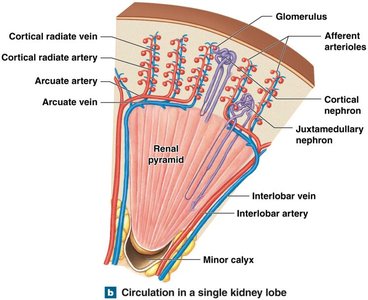
Kidney Lobe: Consists of a renal pyramid, overlying cortex, and adjacent renal columns; each lobe produces urine.
Blood Supply
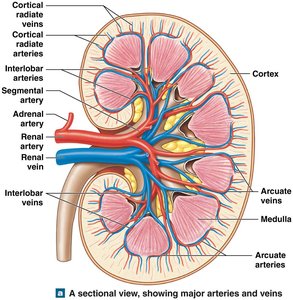
The kidneys receive 20–25% of total cardiac output, approximately 1200 mL of blood per minute. Blood enters each kidney via the renal artery and is distributed through a network of arteries and veins.
Nephrons
Types and Structure
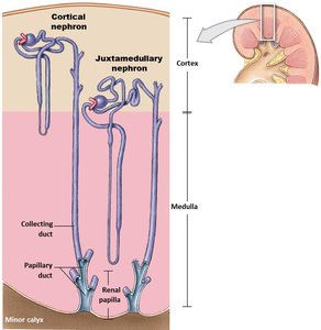
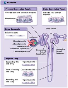
Nephrons are the microscopic functional units of the kidney, responsible for urine formation. Each nephron consists of a renal corpuscle and a renal tubule, which empties into the collecting system.
Cortical Nephrons: 85% of nephrons, located mostly in the superficial cortex.
Juxtamedullary Nephrons: 15% of nephrons, with loops extending deep into the medulla.
Anatomy of a Nephron
Renal Corpuscle: Spherical structure containing the glomerular (Bowman's) capsule and glomerulus (capillary network).
Renal Tubule: Includes the proximal convoluted tubule (PCT), nephron loop (loop of Henle), and distal convoluted tubule (DCT).
Glomerular Filtration
Structure and Function
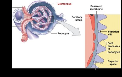
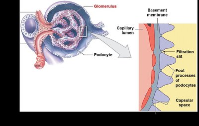
The glomerulus consists of about 50 intertwined capillaries. Blood enters via the afferent arteriole and exits through the efferent arteriole. Podocytes, large cells of the visceral layer, have foot processes (pedicels) that wrap around capillaries, forming filtration slits.
Filtration Membrane: Composed of fenestrated endothelium, basement membrane, and podocyte foot processes.
Filtration Process: Blood pressure forces water and small solutes across the membrane into the capsular space; larger solutes (e.g., plasma proteins) are excluded, producing a protein-free filtrate.
Urine Formation
Basic Processes
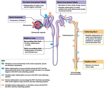
Urine formation involves three main processes: filtration, reabsorption, and secretion. These occur in different segments of the nephron and collecting system.
Filtration: Blood pressure forces water and solutes across glomerular capillary walls.
Reabsorption: Movement of water and solutes (e.g., ions, organic nutrients) from filtrate back into the blood.
Secretion: Transport of solutes from blood into the filtrate for excretion.
Urinary Tract
Ureters
The ureters are paired muscular tubes that transport urine from the kidneys to the urinary bladder. They begin at the renal pelvis, pass over the psoas major, and penetrate the posterior wall of the bladder at an oblique angle. Ureteric orifices are slit-like, preventing backflow during bladder contraction.
Urinary Bladder
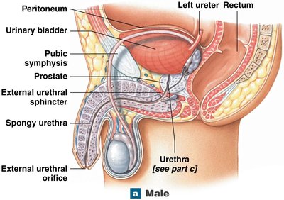
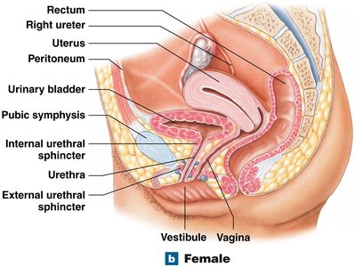
The urinary bladder is a hollow, muscular organ that temporarily stores urine. It can hold up to 1 liter of urine and is stabilized by peritoneal folds and ligamentous bands anchoring it to pelvic and pubic bones.
Urethra
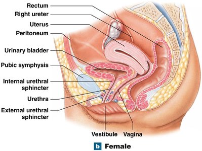
The urethra transports urine from the neck of the bladder to the exterior. Its lamina propria is thick and elastic, and the mucosa has longitudinal folds with mucus-secreting cells in epithelial pockets.
Male Urethra
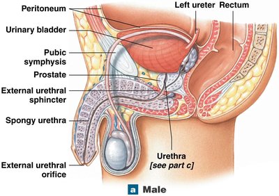
Extends from bladder neck to tip of penis (18–20 cm).
Includes prostatic, membranous, and spongy segments.
Also transports semen in males.
Female Urethra
Short (3–5 cm), extends from bladder to vestibule.
External urethral orifice is near the anterior wall of the vagina.
Summary Table: Main Organs and Functions of the Urinary System
Organ | Main Function |
|---|---|
Kidneys | Produce urine, regulate blood composition |
Ureters | Transport urine to bladder |
Urinary Bladder | Temporary urine storage |
Urethra | Conducts urine to exterior |
Key Terms and Definitions
Urination (Micturition): Process of eliminating urine from the body.
Renal Corpuscle: Structure in nephron where filtration occurs.
Podocyte: Specialized cell in the glomerulus involved in filtration.
Filtration Membrane: Barrier in the renal corpuscle that allows selective passage of substances.
Relevant Equations
Glomerular Filtration Rate (GFR): The rate at which filtrate is produced in the kidneys.
Renal Blood Flow: The volume of blood delivered to the kidneys per unit time.
Additional info: Equations are provided for academic context; actual values depend on physiological parameters.
