 Indietro
Tissue Level of Organization: Structure and Function of Primary Tissues
Guida di studio - Note intelligenti
 Appunti personalizzati basati sui tuoi materiali, ampliati con definizioni chiave, esempi e contesto.
Tissue Level of Organization
Overview of the Four Primary Tissue Types
The human body is composed of four primary tissue types, each with distinct structural and functional characteristics. These tissues are essential for maintaining the integrity, function, and communication within the body.
Epithelial Tissue: Covers exposed surfaces, lines internal passageways, and forms glands. Functions include protection, exchange, and secretion.
Connective Tissue: Fills internal spaces, supports other tissues, transports materials, and stores energy.
Muscle Tissue: Specialized for contraction, enabling movement of the body and its parts.
Nervous Tissue: Transmits electrical signals, facilitating communication between different regions of the body.
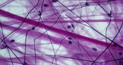
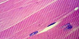
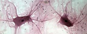
Summary Table: Structure and Function of Primary Tissues
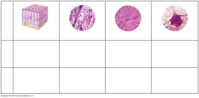
This table summarizes the structural and functional differences among the four primary tissue types.
Tissue | Structure | Function |
|---|---|---|
Epithelial | Sheets of closely packed cells | Protection, exchange, secretion |
Connective | Sparse cells in extracellular matrix | Binding and support of other tissues |
Muscle | Long cells (fibers) with contractile proteins | Movement of body parts |
Nervous | Neurons with branching extensions | Transmission of nerve signals |
Epithelial Tissue
General Characteristics
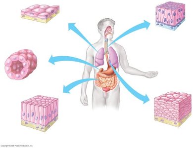
Epithelial tissue lines and covers body surfaces and forms glandular structures. It is avascular (lacks direct blood supply) and has minimal extracellular space, with cells tightly packed together. Epithelial cells are held together by specialized projections on their lateral surfaces.
Lines and covers body surfaces
Forms glandular tissue
Avascular
Cells closely packed
Types of Epithelial Tissue
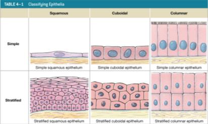
Epithelial tissues are classified based on cell shape and arrangement:
Cell shapes: Squamous (flat), Cuboidal (cube-shaped), Columnar (tall)
Arrangement: Simple (one layer), Stratified (multiple layers)
Surface Modifications
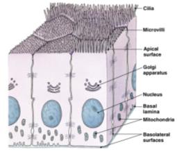
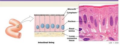
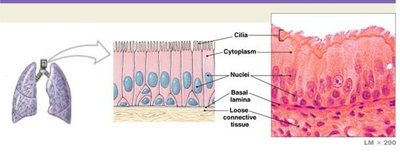
The apical surface of epithelial cells may be modified to form microvilli (increase absorption, common in digestive tract) or cilia (move fluids, common in respiratory tract). The basal layer rests on the basement membrane.
Functions of Epithelial Tissue
Physical protection: Protects underlying tissues from mechanical and chemical stress.
Specialized secretions: Glandular epithelium produces substances for protection and signaling.
Absorption and permeability: Controls movement of substances across the epithelium.
Sensation: Specialized neuroepithelium provides sensory functions (e.g., in eye, ear, nose).
Cell Junctions in Epithelial Tissue
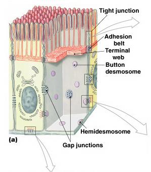
Epithelial cells form bonds with each other and extracellular material through specialized junctions:
Tight junctions: Prevent passage of water and solutes, isolate wastes in the lumen.
Gap junctions: Allow rapid communication via channel proteins (connexons), enable coordinated contractions (e.g., in heart muscle).
Desmosomes: Link opposing plasma membranes via cell adhesion molecules (CAMs), provide strength and flexibility.
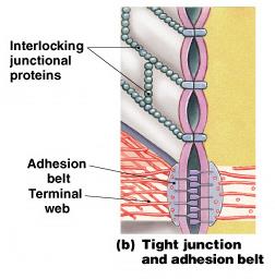
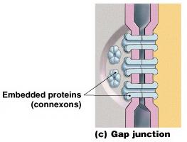
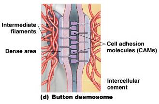
Specific Types of Epithelial Tissue
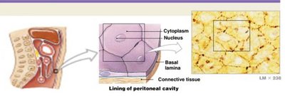
Simple Squamous: Lines ventral body cavities (mesothelium), heart, blood, and lymph vessels (endothelium). Allows easy exchange of materials.
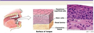
Stratified Squamous: Forms epidermis, lining of mouth and throat. Many cell layers, adapted for protection; keratin adds strength and water resistance.
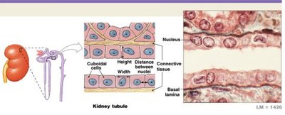
Simple Cuboidal: Found in glands and kidney tubules; adapted for secretion and absorption.
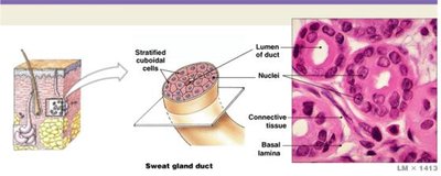
Stratified Cuboidal: Sweat glands and mammary ducts; functions in protection, secretion, and absorption.
Simple Columnar: Lines stomach and digestive tract; functions in protection, secretion, and absorption. Microvilli increase surface area.
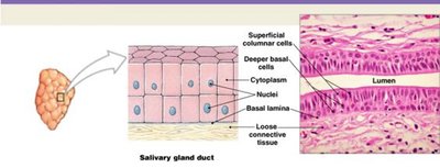
Stratified Columnar: Pharynx, salivary gland ducts, epiglottis, anus; functions in protection.
Pseudostratified: Lines nasal cavity, trachea, and bronchi; functions in protection and secretion. Appears multilayered but is a single layer.
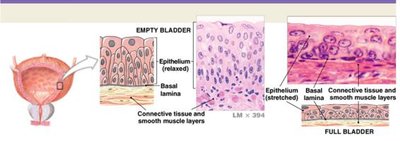
Transitional: Lines hollow organs subject to shape changes (e.g., bladder); many cell layers, adapted for stretching.
Connective Tissue
General Characteristics
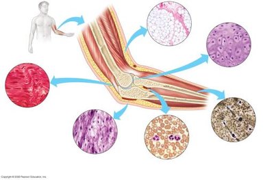
Connective tissue connects epithelium to the rest of the body, provides structure, stores energy, transports materials, and plays a role in protection and tissue repair. It is characterized by more intercellular material than cells and has no direct contact with the environment.
Classification of Connective Tissue
Supportive Connective Tissue: Provides structural strength (cartilage and bone).
Fluid Connective Tissue: Transports materials (blood and lymph).
Connective Tissue Proper: Connects and protects; includes loose and dense connective tissues.
Supportive Connective Tissue: Cartilage
Cartilage consists of cells in a gel-like matrix, providing shock absorption and protection. It is avascular and nourished by the perichondrium, which has an outer fibrous layer for strength and an inner cellular layer for growth and maintenance.
Elastic Cartilage: Tightly packed elastic fibers, flexible support, found in external ear and epiglottis.
Hyaline Cartilage: Reduces friction in joints, connects ribs to sternum, found at ends of moving bones.
Fibrocartilage: Resists compression, prevents bone-to-bone contact, dense collagen fibers, found in knee joints, intervertebral discs, and pubic bones.
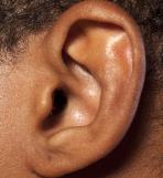
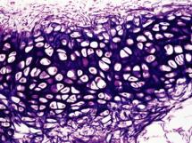
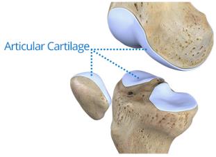
Supportive Connective Tissue: Bone
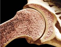
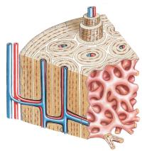
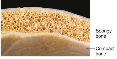
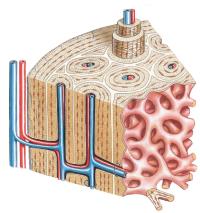
Bone (osseous tissue) is strong due to calcified Ca2+ salt deposits and flexible collagen fibers. Osteocytes are arranged around central canals, and canaliculi provide access to blood supply. The periosteum covers bone surfaces and consists of fibrous and cellular layers.
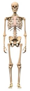
Fluid Connective Tissue: Blood and Lymph
Blood and lymph are fluid connective tissues with a watery matrix. The internal environment of tissues is comprised of interstitial fluid, which serves as a medium for transfer of solutes and wastes between blood, lymph vessels, and cells.
Connective Tissue Proper
Connective tissue proper includes loose and dense connective tissues. Loose connective tissue fills spaces between organs, cushions and stabilizes cells, and surrounds blood vessels. Adipose tissue provides padding and insulation, and dense connective tissue (collagenous) provides strength and elasticity.
Membranes
Types of Membranes
Membranes are physical barriers that line or cover portions of the body. They consist of an epithelium supported by connective tissue and serve protective and friction-reducing functions.
Mucous Membranes: Line passageways with external connections (digestive, respiratory, urinary, reproductive tracts).
Serous Membranes: Line cavities not open to the outside; reduce friction via transudate.
Cutaneous Membrane: The skin; covers the body surface, thick, waterproof, and dry.
Synovial Membranes: Line joint cavities, produce synovial fluid for lubrication, protect ends of moving bones.
Functions and Locations of Membranes
Protection: Physical and chemical barrier.
Serous membranes: Reduce friction between contacting structures.
Location: Line body cavities (viscera), cover organs, inner lining of vascular system (heart, arteries, veins, lymphatic vessels).
Mucous Membranes
Mucosae line passageways that have external connections, such as the digestive, respiratory, urinary, and reproductive tracts.
Serous Membranes
Serous membranes line cavities not open to the outside and cover organs within these cavities. The pleural membrane covers the lungs, the peritoneum covers abdominal organs, and the pericardium covers the heart.
Cutaneous Membrane
The cutaneous membrane, or skin, covers the body surface and consists of stratified squamous epithelium, areolar tissue, and dense irregular connective tissue.
Synovial Membranes
Synovial membranes line articulating joint cavities, produce synovial fluid for lubrication, and protect the ends of moving bones.
