 Indietro
Tissue Level Organization: Structure and Function of Human Tissues
Guida di studio - Note intelligenti
 Appunti personalizzati basati sui tuoi materiali, ampliati con definizioni chiave, esempi e contesto.
Tissue Level Organization
Introduction to Tissues
Tissues are collections of specialized cells and cell products that perform specific functions. The study of tissues is known as histology. Tissues combine to form organs, which then interact in organ systems to carry out complex functions in the body.
Four major types of tissues: Epithelial, Connective, Muscle, Nervous
Role: Each tissue type has distinct structural and functional characteristics.
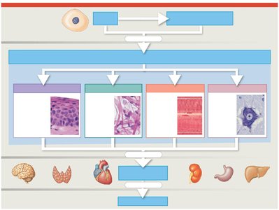
Overview of Tissue Types
Epithelial Tissue: Covers exposed surfaces, lines internal passageways, and forms glands.
Connective Tissue: Fills internal spaces, provides structural support, and stores energy.
Muscle Tissue: Contracts to produce movement.
Nervous Tissue: Propagates electrical impulses and carries information.
Epithelial Tissue
Structure and Functions
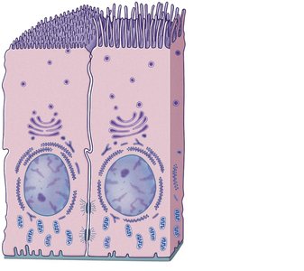
Epithelial tissue includes layers of cells covering internal or external surfaces and glands that produce fluid secretions. It serves several essential functions:
Physical protection
Control of permeability
Provision of sensation
Production of specialized secretions
Integrity and Intercellular Connections
The integrity of epithelia is maintained by intercellular connections, attachment to the basement membrane, and ongoing maintenance and repair.
Cell junctions: Specialized structures that connect cells or attach them to extracellular material.
Types of cell junctions: Gap junctions, Tight junctions, Desmosomes (spot and hemidesmosomes)
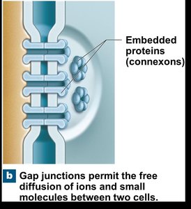
Gap Junctions
Gap junctions allow rapid communication between cells by permitting the free diffusion of ions and small molecules. They are formed by interlocking transmembrane proteins called connexons.
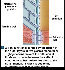
Tight Junctions
Tight junctions are formed by the fusion of the outer layers of two plasma membranes. They prevent the passage of water and solutes, maintaining compartmentalization within tissues.
Desmosomes
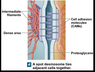
Desmosomes are cell junctions that tie adjacent cells together, allowing tissues to withstand mechanical stress. Spot desmosomes connect cells, while hemidesmosomes attach cells to the basement membrane.
Hemidesmosomes and Basement Membrane
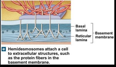
Hemidesmosomes attach a cell to extracellular structures, such as the protein fibers in the basement membrane. The basement membrane consists of the basal lamina (closest to the epithelium) and the reticular lamina (provides strength).
Classification of Epithelia
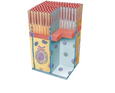
Epithelia are classified based on cell shape and the number of cell layers:
Shape: Squamous (thin, flat), Cuboidal (square), Columnar (tall, slender)
Layers: Simple (single layer), Stratified (multiple layers)
Glandular Epithelia
Endocrine glands: Release hormones directly into the bloodstream (no ducts).
Exocrine glands: Discharge secretions through ducts onto epithelial surfaces.
Connective Tissue
Components and Functions
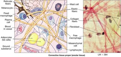
Connective tissues consist of specialized cells, extracellular protein fibers, and ground substance. The matrix (fibers and ground substance) determines the tissue's specialized function.
Functions: Structural framework, transport, protection, support, energy storage, defense
Categories of Connective Tissue
Connective tissue proper: Connects and protects (loose and dense types)
Fluid connective tissues: Blood and lymph (transport)
Supporting connective tissues: Cartilage and bone (structural strength)
Connective Tissue Proper
Loose connective tissue has more ground substance and fewer fibers (e.g., adipose tissue), while dense connective tissue has more fibers and less ground substance (e.g., tendons).
Fluid Connective Tissues
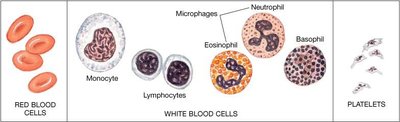
Blood consists of formed elements (red blood cells, white blood cells, platelets) suspended in plasma. Lymph is interstitial fluid entering lymphatic vessels.
Supporting Connective Tissues
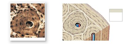
Cartilage is a firm gel containing chondroitin sulfate, with cells called chondrocytes in lacunae. Bone (osseous tissue) is rigid due to calcium salts and flexible collagen fibers. Osteocytes lie in lacunae and are arranged around central canals.
Muscle Tissue
Types and Functions
Muscle tissue is specialized for contraction and movement. There are three types:
Skeletal muscle: Large, striated, voluntary muscles responsible for body movement
Cardiac muscle: Striated, involuntary muscle found only in the heart
Smooth muscle: Nonstriated, involuntary muscle found in walls of hollow organs
Skeletal Muscle Tissue
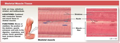
Skeletal muscle fibers are long, cylindrical, striated, and multinucleate. They move or stabilize the position of the skeleton and guard entrances and exits.
Cardiac Muscle Tissue
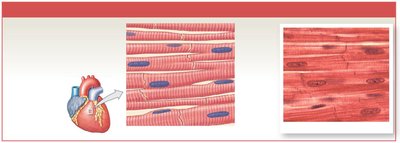
Cardiac muscle cells are short, branched, and interconnected by intercalated discs. They circulate blood and maintain blood pressure.
Smooth Muscle Tissue
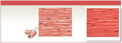
Smooth muscle cells are short, spindle-shaped, and nonstriated. They move food, urine, and reproductive tract secretions, and regulate the diameter of blood vessels.
Nervous Tissue
Structure and Function
Nervous tissue is specialized for conducting electrical impulses and is concentrated in the brain and spinal cord. It consists of two main cell types:
Neurons: Conduct electrical impulses
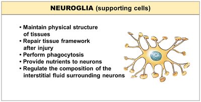
Neuroglia: Support, protect, and nourish neurons
Neuroglia (Supporting Cells)
Maintain physical structure of tissues
Repair tissue framework after injury
Perform phagocytosis
Provide nutrients to neurons
Regulate the composition of interstitial fluid
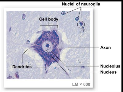
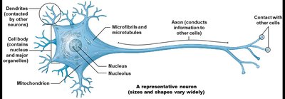
Parts of a Neuron
Cell body: Contains nucleus and nucleolus
Dendrites: Receive incoming signals
Axon: Carries outgoing electrical signals
Summary Table: Major Tissue Types
Tissue Type | Main Function | Key Features |
|---|---|---|
Epithelial | Protection, secretion, absorption | Cell layers, polarity, junctions |
Connective | Support, transport, defense | Matrix, fibers, cells |
Muscle | Movement | Contractile proteins, cell shape |
Nervous | Communication | Neurons, neuroglia |
Example: The heart contains muscle tissue (cardiac), connective tissue (support), epithelial tissue (lining), and nervous tissue (regulation).
Additional info: Academic context was added to clarify tissue functions, cell junctions, and classification. All images included are directly relevant to the explanation of their adjacent paragraphs.
